بررسی ارزش تشخیصی یافته های سی تی اسکن درتشخیص نکروز ترانسمورال روده در بیماران با ایسکمی مزانتریک وریدی مراجعه کننده به بیمارستان امام خمینی بین سال های 1392 تا ۱۳۹۸

Background: Acute mesenteric ischemia is an emergent condition needing timely and appropriate diagnosis and management. This study aimed to determine the clinical, laboratory and computed tomography (CT) findings that indicate transmural necrosis in acute mesenteric venous thrombosis patients.
Methods: We used the electronic records and multi-detector CT scans of the patients hospitalized between 2014 and 2021 with the diagnosis of acute mesenteric ischemia due to venous thrombosis. We compared multiple clinical and radiological findings between the patients with transmural necrosis on laparotomy and those who were discharged from the hospital.
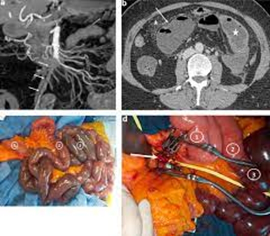
Results: Of 51 patients, 18 underwent laparotomy, and among them, 13 had transmural necrosis, whereas five patients had normal or ischemic bowel. Thirty-three completed conservative therapy. Thirty-two patients had bowel wall thickening on CT scan, and the other 19 patients only had acute mesenteric venous thrombosis and abdominal pain without bowel wall thickening on on-admission CT scan. Among the laboratory findings, the mean level of creatinine, urea, C-reactive protein, lactate dehydrogenase, white blood cells, and the percentage of neutrophils was higher in the transmural necrosis group. Comparing the CT scan findings in all patients, the presence of bowel wall thickening(100% versus 50.0% of the cases, P- value=0.001), adjacent focal mesenteric haziness (92.3% versus 26.3%, P-value<0.001), foci with decreased enhancement (75.0% versus 21.6%, P-value=0.001), pneumatosis intestinalis (23.1% versus 0.0%, P-value=0.01), and subendothelial lucent line in the bowel wall (46.2% versus 7.9%, P-value=0.005) were significantly more seen in transmural necrosis cases. When considering CT findings to predict transmural necrosis in the 32 patients with abnormal bowel wall thickening, the length of the abnormal loop (87.3 ± 82.8 versus 32.4 ± 40.1, P-value=0.01), the diameter of the thickened loop (32.9 ± 7.8 versus 27.5 ± 5.1, P-value=0.02) and proximal bowel (30.9 ± 11.1 versus 24.0 ± 6.9, P-value=0.04), and adjacent focal mesenteric haziness (92.3% versus 52.6%, P-value=0.02) were statistically significantly higher in those with transmural necrosis.). Among patients with abnormal bowel wall thickening, Cr, urea, and CRP among laboratory markers, and amid clinical findings, obstipation was higher in patients with transmural necrosis.
Conclusion: Phcisysions can use laboratory and radiological findings to differentiate transmural necrosis from ischemia in cases with acute mesenteric venous thrombosis.
Keywords: Necrosis, Mesenteric Ischemia, Computed Tomography, Diagnostic Imaging.


ارسال نظر