

Sonographic skin features and shear wave elastography in distinguishing active from inactive morphea lesions: A case-control study
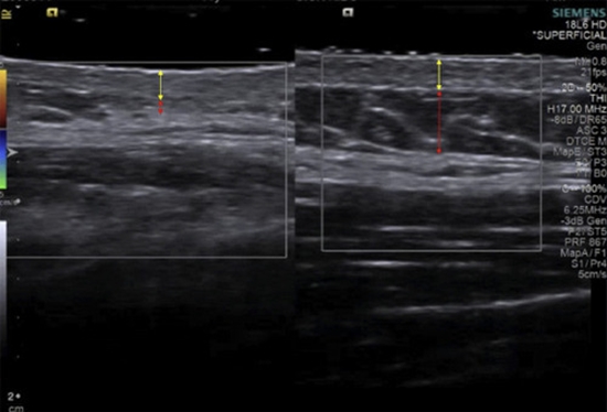
Morphea (localized scleroderma) is a connective tissue disorder characterized by dermal stiffness, thickening, erythema, and pigmentation abnormalities, often varying with the degree of inflammation and fibrosis.1 Precise lesion activity is crucial for tailoring optimal treatment plans. Traditional diagnostic methods like skin biopsy are invasive and may not effectively capture the dynamic progression of the disease.2 Noninvasive imaging techniques, including ultrasound (US) and shear wave elastography (SWE), present promising alternatives for diagnosis and monitoring by assessing vascular changes and skin stiffness.1,3 This case-control study evaluates the effectiveness of US and SWE in distinguishing between active and inactive morphea lesions, aiming to improve the accuracy of morphea management.


ارسال نظر