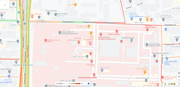
فعالیت های مرکز تحقیقات رادیولوژی دانشگاه علوم پزشکی تهران در دوران همه‌گیری کووید-۱۹

همه‌گیری کووید-۱۹ و نقش مرکز تحقیقات رادیولوژی

در طول همه‌گیری کووید-۱۹، مرکز تحقیقات رادیولوژی دانشگاه علوم پزشکی تهران، علی‌رغم چالش‌های ناشی از افزایش تقاضا برای سی‌تی اسکن بیماران کووید-۱۹ و لزوم رعایت پروتکل‌های بهداشتی سختگیرانه، به فعالیت‌های تحقیقاتی خود ادامه داد. این مرکز نقشی حیاتی در بهبود تکنیک‌های تصویربرداری پزشکی، بهینه‌سازی پروتکل‌های تشخیصی و توسعه فناوری‌های مرتبط با تجزیه و تحلیل تصاویر سی‌تی اسکن ایفا کرد.

اعضای هیئت علمی، محققان و کارکنان ضمن تضمین ایمنی و رفاه متخصصان مراقبت‌های بهداشتی و بیماران، فعالانه در تجزیه و تحلیل داده‌های تصویربرداری، توسعه الگوریتم‌های هوش مصنوعی برای تشخیص سریع‌تر بیماری، انتشار مقالات علمی و ارائه یافته‌های خود در کنفرانس‌های بین‌المللی شرکت داشتند. این تلاش‌ها با هدف افزایش دقت تشخیصی، بهبود فرآیندهای درمان و کاهش فشار بر سیستم‌های مراقبت‌های بهداشتی انجام شد.

دستاوردهای کلیدی مرکز تحقیقات رادیولوژی در طول همه‌گیری کووید-۱۹

  • توسعه و بهینه‌سازی پروتکل‌های تصویربرداری ریه: برای تشخیص سریع‌تر و دقیق‌تر کووید-۱۹
  • پیاده‌سازی فناوری‌های هوش مصنوعی: برای تجزیه و تحلیل خودکار سی‌تی اسکن و کاهش زمان تشخیص
  • انتشار مقالات تحقیقاتی با تأثیر بالا: در مورد روش‌های تصویربرداری نوآورانه برای بیماران کووید-۱۹
  • مشارکت در پروژه‌های ملی و بین‌المللی: متمرکز بر تشخیص کووید-۱۹ و مدیریت بیماران

این مرکز همچنان به پیشبرد تحقیقات در زمینه تصویربرداری پزشکی اختصاص دارد و به عنوان یک مؤسسه علمی پیشرو در بهبود کیفیت خدمات تشخیصی و درمانی به مشارکت خود ادامه می‌دهد.

© مرکز تحقیقات رادیولوژی دانشگاه علوم پزشکی تهران

  • تاریخ انتشار : 1403/11/29 - 13:34
  • number of visits : 33
  • زمان مطالعه : 1 دقیقه

Role of chest CT scan in patients with preexisting cancer and COVID-19 pneumonia

Role of chest CT scan in patients with preexisting cancer and COVID-19 pneumonia {faces}

Background: Detection of COVID-19 in cancer patients is challenging due to probable preexisting pulmonary infiltration caused by many infectious and non-infectious etiologies. We evaluated chest CT scan findings of COVID-19 pneumonia in cancer patients and explored its prognostic role in mortality.

Methods: We studied 266 COVID-19 patients with a history of cancer diagnosis between 2020 and 2022. Chest CT images were reported based on Radiological Society of North America (RSNA) structural report and the CT score and pattern of involvement were noted. We used multivariate logistic regression models to determine the association between CT scan findings and mortality of the cancer COVID-19 patients.

Results: The mean age was 56.48 (± 18.59), and 53% were men. Gastrointestinal (29.3%), hematologic (26.3%), and breast (10.5%) cancers were the most frequent types of cancer. The prevalence of atypical or indeterminate findings in the chest CT was 42.8%. Most radiologic findings were consolidation mixed with ground-glass opacity (44.4%), pleural effusion (33.5%), and pure ground-glass opacity (19.5%). The risk of death was higher among those who had typical chest CT for COVID-19 (OR 3.47; 95% CI 1.14-8.98) and those who had a severity of score higher than 18 (OR 1.89; 95% CI 1.07-3.34). Also, presence of consolidation (P value 0.040), pleural effusion (P value 0.000), centrilobular nodules (P value 0.013), and architectural distortion (P value 0.005) were associated with a poorer prognosis.

Conclusion: Less than half of COVID-19 patients with a history of cancer had typical imaging features of COVID-19. Radiologists should be aware of atypical, rare, or subtle chest CT findings in patients with pre-existing cancer.

  • Article_DOI : 10.1186/s12880-023-00984-w
  • نویسندگان : masoumeh gity,faezeh khorasanizadeh,soori kaviani,shadi salamroudi ,monireh sadat seyyedsalehi
  • گروه خبر : پژوهش,research article,covid19
  • کد خبر : 290714
مدیر سیستم
تهیه کننده:

مدیر سیستم

0 نظر برای این مطلب وجود دارد

ارسال نظر

نظر خود را وارد نمایید:

متن درون تصویر را در جعبه متن زیر وارد نمائید *
متن مورد نظر خود را جستجو کنید
تنظیمات پس زمینه
Close menu