.jpg)


Role of chest CT scan in patients with preexisting cancer and COVID-19 pneumonia
Background: Detection of COVID-19 in cancer patients is challenging due to probable preexisting pulmonary infiltration caused by many infectious and non-infectious etiologies. We evaluated chest CT scan findings of COVID-19 pneumonia in cancer patients and explored its prognostic role in mortality.
Methods: We studied 266 COVID-19 patients with a history of cancer diagnosis between 2020 and 2022. Chest CT images were reported based on Radiological Society of North America (RSNA) structural report and the CT score and pattern of involvement were noted. We used multivariate logistic regression models to determine the association between CT scan findings and mortality of the cancer COVID-19 patients.
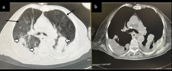
Results: The mean age was 56.48 (± 18.59), and 53% were men. Gastrointestinal (29.3%), hematologic (26.3%), and breast (10.5%) cancers were the most frequent types of cancer. The prevalence of atypical or indeterminate findings in the chest CT was 42.8%. Most radiologic findings were consolidation mixed with ground-glass opacity (44.4%), pleural effusion (33.5%), and pure ground-glass opacity (19.5%). The risk of death was higher among those who had typical chest CT for COVID-19 (OR 3.47; 95% CI 1.14-8.98) and those who had a severity of score higher than 18 (OR 1.89; 95% CI 1.07-3.34). Also, presence of consolidation (P value 0.040), pleural effusion (P value 0.000), centrilobular nodules (P value 0.013), and architectural distortion (P value 0.005) were associated with a poorer prognosis.
Conclusion: Less than half of COVID-19 patients with a history of cancer had typical imaging features of COVID-19. Radiologists should be aware of atypical, rare, or subtle chest CT findings in patients with pre-existing cancer.

