.jpg)


Ultrasound and magnetic resonance imaging features of fetal intracranial cystic lesions: A pictorial essay
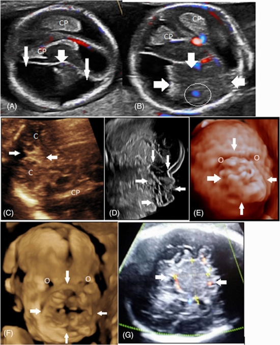
This pictorial essay focuses on ultrasound (US) and magnetic resonance imaging (MRI) features of fetal intracranial cysts. Intracranial cysts are common findings in prenatal imaging, and if great attention is paid to their size, location, and imaging features, they can be diagnosed accurately. They are usually detected by fetal ultrasound exams. However, when ultrasound data on cystic lesion characteristics is insufficient, MRI and fetal neurosonogram are the best options for detecting other associated anomalies. The prognosis is highly dependent on their location and whether they are associated with other fetal anomalies.

