3T magnetic resonance spectroscopy as a powerful diagnostic modality for assessment of thyroid nodules

Objective
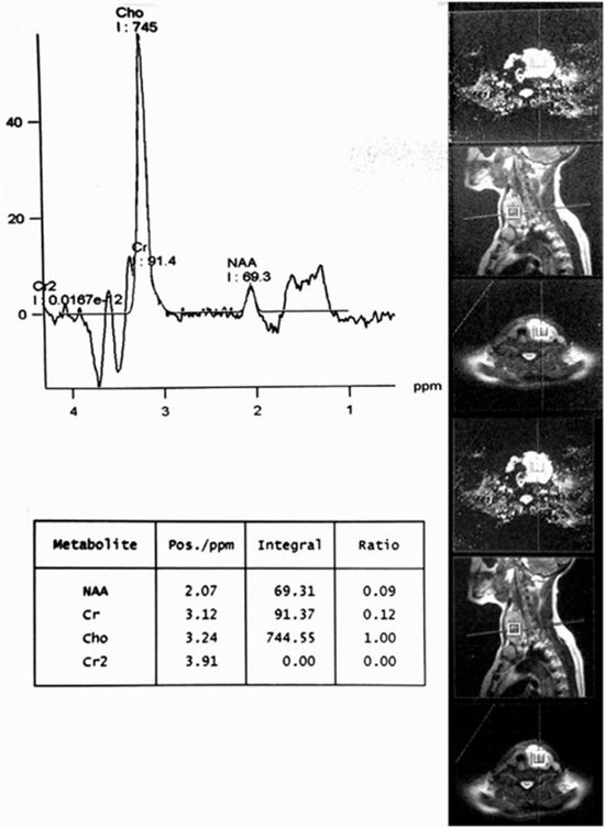
Magnetic resonance spectroscopy (MRS) is a powerful tool for structural studies of chemical compounds and biomolecules and also documented promising findings as a potential imaging technology in thyroid oncology. This prospective study was to ascertain the clinical significance of 3 Tesla MRS in the evaluation of patients with thyroid nodules (TNs) as an ancillary diagnostic technique for thyroid carcinoma.
Materials and methods
Magnetic resonance spectroscopy at 3T at echo- times (TEs) 136 and 270 ms was carried out on 15 patients with total number of 32 TNs larger than 1 cm3, which all were surgically resected. Choline (Chol) to creatine (Cr) ratio was assessed at 136 and 270 TEs on each nodule and a receiver operating characteristic (ROC) curve was used to determine optimal cut-off point. The findings were compared with histopathology of thyroid specimens.
Results
There were 23 benign and 9 malignant lesions (7 papillary and 2 follicular thyroid carcinomas). The mean values of Chol/Cr at 136 and 270 TEs was 2.28 ± 3.65 and 1.52 ± 1.67 respectively and the difference between benign and malignant nodules was only significant at 136 TEs. The study revealed that Chol/ Cr ratio cut-off point of 2.5 best correlates with histopathology results (sensitivity = 75%; specificity = 100%; PPV = 100%; NPV= 92%).
Conclusion
This preliminary study showed that 3T magnetic resonance spectroscopy might be a specific modality for the evaluation of thyroid nodules in differentiation of benign from malignant thyroid tissue. However, a larger series would give much greater confidence that this state-of-the-art technology will worth pursuing in clinical practice


ارسال به دوستان