بررسی حساسیت و اختصاصیت MRI پرفیوژن در درج بندی تومورهای گلیال مغز
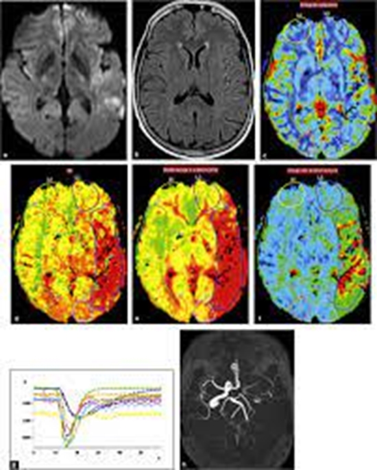
تعداد 35 بیمار مراجعه کننده به درمانگاه جراحی اعصاب بیمارستان امام خمینی مبتلا به گلیوم مغزی بر اساس یافته های تصویر برداری مرسوم (T1,T2,T1+contrast)که کاندید جراحی بودند تحت تصویر برداری باپروتکل perfusion MRI قرار گرفتند.میزان حجم خون مغز در منطقه تومورال(CBVt),حجم خون مغزدر ناحیه ادم(CBVe)و ماده سفید نرمال سمت مقابل (CBVwm) طبق نرم افزار MATLAB محاسبه شد.میزان نسبی حجم خون مغز ناحیه تومورال (rCBVt)با تقسیم مقدار CBVt برCBVwm ومیزان نسبی حجم خون ناحیه ادم اطراف تومور(rCBVe)با تقسیم CBVeبرCBVwm سمت مقابل محاسبه شد.جواب های rCBVt,rCBVeبا گرید پاتولوژی تومور بر اساس گریدWHO مقایسه شد.اقدامات فوق برای ی جریان خون مغزی نسبی(rCBF) در ناحیه تومورال(rCBFt)و ادم اطراف آن(rCBFe) نیز صورت گرفت.

هدف:
در این مطالعه هدف ما مقایسه حجم خون مغزی (CBV) و جریان خون مغزی (CBF) در درجهبندی گلیوما بود.
مواد و روشها:
مجموعاً ۳۵ بیمار با یافتههای MRI متداول مشکوک به تومور گلیال تحت MRI پرفیوژن با دستگاه 3T قرار گرفتند. شاخصهای rCBV تومور (rCBVt)، rCBV ادم (rCBVe)، rCBF تومور (rCBFt) و rCBF ادم (rCBFe) محاسبه شدند.
یافتهها:
میانگین سنی بیماران ۴۴.۵±۱۹ سال (دامنه ۱۲ تا ۸۴ سال) بود. ۲۰ بیمار (۵۷٪) مرد بودند. در مجموع ۲۰ بیمار دارای تومور با درجه بالا (درجه ۳ و ۴) بودند. نتایج نشان داد که rCBV بهتنهایی ارزش تشخیصی کافی برای تمایز بین تومورهای درجه پایین و بالا ندارد.
نتیجهگیری:
اگرچه مقایسه rCBV و rCBF نشان داد میانگین این مقادیر بین تومورهای درجه بالا و پایین از نظر آماری متفاوت است، اما rCBF میتواند کاربردیتر از rCBV در درجهبندی گلیوما باشد.


ارسال نظر