بررسی همبستگی یافته های MRI و CT اسکن در بیماران مبتلا به سیستیک فیبروزیس در مرکز طبی کودکان شهر تهران طی سال های ۱۳۹۸ و ۱۳۹۹

Abstract
Objective: To evaluate whether magnetic resonance imaging (MRI) is effective as computed tomography (CT) in determining functional pulmonary changes in patients with cystic fibrosis (CF)
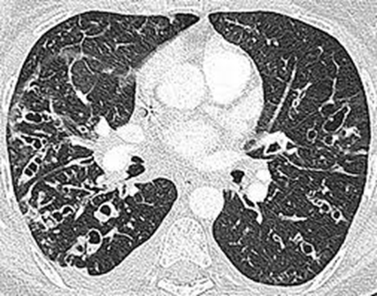
Methods: Twenty-three patients (12 males) aging between 1 to 20 years, CF disease of whom was confirmed by sweat test, were studied for 2 years. Patients were evaluated using lung CT scan, and one-week later chest MRI, without contrast injection. Consistency of 222 variables was tested using the Bhalla scoring system for the findings of CT and MRI
Results: There was a strong correlation between Bhalla Chest CT and MRI Scores (R = 0.97, p<0.0001). The mean and median Helbich-Bhalla score for CT were 16.87 and 16 (range: 7–25) and those of MRI were 17.48 and 17 (range: 8-25). The mean and median difference were 0.61 and 1 points, respectively. Chest CT and MRI were strongly correlated and showed a high level of agreement to diagnose all variables studied, including the severity and extent of bronchiectasis (ICC: 98.8% and 97.6%; r = 0.988 and 0.977, respectively), peribronchial thickening (ICC: 81.4%; r = 0.815), sacculations, generalities of the bronchial division involved, and number of bubbles (ICC: 100%; r = 1), emphysema (ICC: 67.3%; r = 1) and collapse/ consolidation (ICC: 89.1%; r = 0.890)
Conclusion: These results showed that none of the relevant findings were missed by MR imaging and MRI can be a safer yet reliable alternative to CT to follow pulmonary changes in CF patients
Keywords: Cystic fibrosis, MRI, CT scan, Pulmonary change


ارسال نظر