Advanced Diagnostic & Interventional Radiology Research Center | Liver Hemangioma Using Trans-Catheter Arterial Embolization

Advanced Diagnostic & Interventional Radiology Research Center | Liver Hemangioma Using Trans-Catheter Arterial Embolization
| Jan 3 2026
logo

Advanced Diagnostic & Interventional Radiology Research Center

COVID-19 pandemic 

During the COVID-19 pandemic, the Radiology Research Center at Tehran University of Medical Sciences continued its research activities despite the challenges posed by the increased demand for CT scans of COVID-19 patients and the necessity of adhering to strict health protocols. This center played a crucial role in improving medical imaging techniques, optimizing diagnostic protocols, and advancing technologies related to CT scan image analysis.

Faculty members, researchers, and staff remained committed to ensuring the safety and well-being of healthcare professionals and patients while actively engaging in imaging data analysis, developing artificial intelligence algorithms for faster disease detection, publishing scientific articles, and presenting their findings at international conferences. These efforts aimed to enhance diagnostic accuracy, improve treatment processes, and alleviate pressure on healthcare systems.


Key achievements of the Radiology Research Center during the COVID-19 pandemic include:


✔️ Development and optimization of lung imaging protocols for faster and more accurate COVID-19 diagnosis
✔️ Implementation of artificial intelligence technologies for automated CT scan analysis and reduced diagnosis time
✔️ Publication of high-impact research articles on innovative imaging methods for COVID-19 patients
✔️ Participation in national and international projects focused on COVID-19 diagnosis and patient management

The center remains dedicated to advancing research in medical imaging and continues to contribute as a leading scientific institution in improving the quality of diagnostic and therapeutic services.


Some of the center's significant achievements during the pandemic include:


  • Release Date : Feb 27 2024 - 14:15
  • : 20
  • Study time : 1 minute(s)

Management of Liver Hemangioma Using Trans-Catheter Arterial Embolization

Hemangioma, a congenital vascular malformation, is the most common benign liver lesion that is usually remain stable subsequently requiring not treatment; however, complications such as abdominal pain or fullness, coagulation disturbances, and inflammatory syndrome may occur, demanding a specific treatment of hemangioma.

Management of Liver Hemangioma {faces}

Background:

Hemangioma, a congenital vascular malformation, is the most common benign liver lesion that is usually remain stable subsequently requiring not treatment; however, complications such as abdominal pain or fullness, coagulation disturbances, and inflammatory syndrome may occur, demanding a specific treatment of hemangioma.

Objectives:

To assess the safety, feasibility and efficacy of trans-catheter arterial embolization (TAE) for the treatment of Liver hemangioma

Patients and Methods:

TAE was performed on 20 patients with liver hemangioma. The embolic agent used was polyvinyl alcohol (PVA) particles (300-400 micron, Jonson and Johnson Cordis, USA). All patients were followed up for 6 months. Imaging was carried out and patients were also evaluated symptomatically through telephone interview by a physician.

Results:

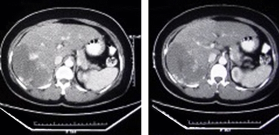
Twenty patients aged from 21 to 63 years (mean: 46.8, SD: 10.26) were included in this study. Post embolization syndrome, including abdominal pain, fever, and leukocytosis occurred in one patient 1 week after TAE and lasted for 3 days. No serious adverse event and TAE-related death was observed. None of the patient underwent another intervention including surgery. During follow up interval, decreased episode of abdominal pain was documented in all patients who had pain. Tumor enlargement was also stopped during the follow up. The average diameter of tumors was 97.00 mm (range: 25-200 SD: 47.85) and 88.95 mm (range: 23-195 SD: 43.27) before and after embolization, respectively. Comparison of images before and after TAE revealed statistically significant decrease in the size of lesion (P value: 0.004, t: 3.31).

Conclusions:

Our findings indicate that TAE is a safe and efficient procedure for the treatment of liver hemangioma. Further studies with larger sample sizes are required to support therapeutic effects of TAE.

  • Article_DOI : doi.org/10.5812/hepatmon.25788
  • Author(s) : kavous firouznia ,sima hosseinverdi
  • News Group : research,research article
  • News Code : 278489
مدیر سایت
Author:

مدیر سایت

Enter your desired term to search
Theme settings