


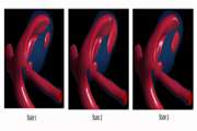
Effects of Variations of Flow and Heart Rate on Intra-Aneurysmal Hemodynamics in a Ruptured Internal Carotid Artery Aneurysm During Exercise
This study aimed to investigate the effects of variations in heart rate and internal carotid artery (ICA) flow rate on intra-aneurysmal hemodynamics, in an ICA aneurysm, by using computational fluid dynamics
Coil Embolization Of Intracranial Aneurysms: A Six-Month Follow-Up Study
It has been established that presence of lean umbilical cord with reduced Wharton's jelly in sonographic scans is a fetal marker for risk of small for gestational age at birth. With improvement of ultrasound techniques, more studies have been investigating the alterations of the umbilical cord on pregnancy outcomes
Comparison of endovascular coiling and surgical clipping for the treatment of intracranial aneurysms: A prospective study
The aim of this study was to compare the interventional outcomes between two groups of patients, one treated with endovascular coiling and the other treated with surgical clipping
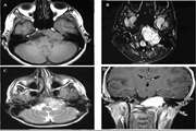
Skull Base Aneurysmal Bone Cyst Presented with Foramen Jugular Syndrome and Multi-Osseous Involvement
Aneurysmal bone cyst (ABC) is an expansile bone lesion that usually involves the long bones
The Prospective Profile of Endovascular Coiling of Cerebral Aneurysms: The Impact of Clinical Presentation, Procedural Results, Aneurysm Rupture and Location
Selection of voxels with the guide of chemical shift imaging map yields to 100% diagnostic sensitivity. If not accessible, the use of the union of peripheral and central voxels enhances the sensitivity when compared to usage of peripheral or central voxels solely.
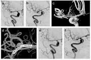
Traumatic Dissecting Posterior Cerebral Artery Aneurysm: A Case Report and Review of the Literature
Dissecting posterior cerebral artery (PCA) aneurysms are among rare cerebrovascular malformations accounting for 2% to 6% of all aneurysms.
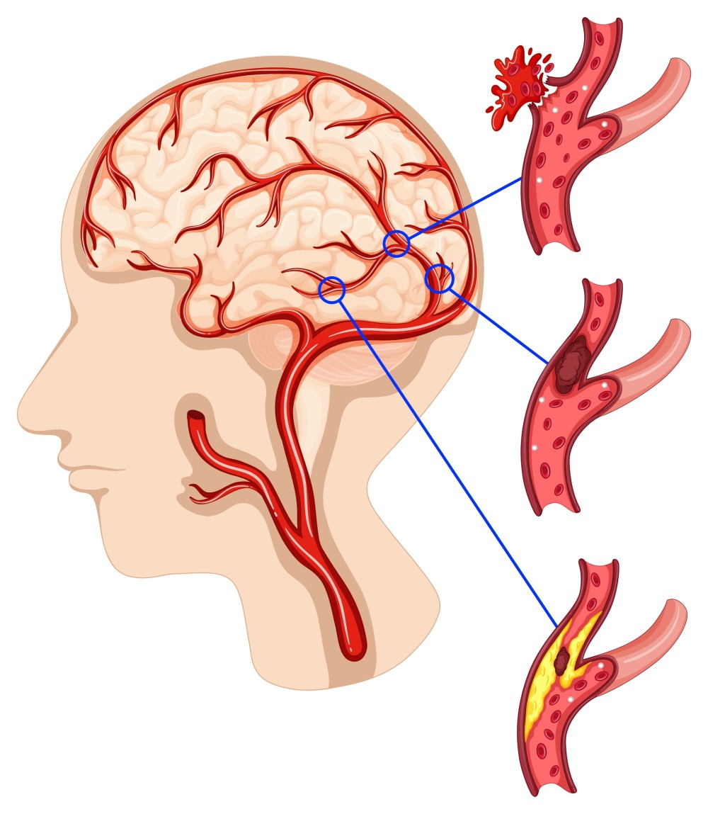
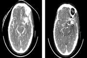
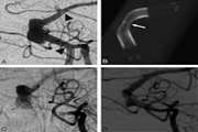
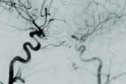
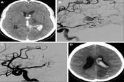
Evaluation of angiographic findings in spontaneous aneurysmal subarachnoid hemorrhage patients
This paper describes the angiographic findings of spontaneous aneurysmal subarachnoid hemorrhage (SAH) patients, including frequency, anatomic location and multiplicity of cerebral aneurysms
