Advanced Diagnostic & Interventional Radiology Research Center | Optimal method for early detection of cardiac disorders

Advanced Diagnostic & Interventional Radiology Research Center | Optimal method for early detection of cardiac disorders
| Jan 2 2026
logo

Advanced Diagnostic & Interventional Radiology Research Center

COVID-19 pandemic 

During the COVID-19 pandemic, the Radiology Research Center at Tehran University of Medical Sciences continued its research activities despite the challenges posed by the increased demand for CT scans of COVID-19 patients and the necessity of adhering to strict health protocols. This center played a crucial role in improving medical imaging techniques, optimizing diagnostic protocols, and advancing technologies related to CT scan image analysis.

Faculty members, researchers, and staff remained committed to ensuring the safety and well-being of healthcare professionals and patients while actively engaging in imaging data analysis, developing artificial intelligence algorithms for faster disease detection, publishing scientific articles, and presenting their findings at international conferences. These efforts aimed to enhance diagnostic accuracy, improve treatment processes, and alleviate pressure on healthcare systems.


Key achievements of the Radiology Research Center during the COVID-19 pandemic include:


✔️ Development and optimization of lung imaging protocols for faster and more accurate COVID-19 diagnosis
✔️ Implementation of artificial intelligence technologies for automated CT scan analysis and reduced diagnosis time
✔️ Publication of high-impact research articles on innovative imaging methods for COVID-19 patients
✔️ Participation in national and international projects focused on COVID-19 diagnosis and patient management

The center remains dedicated to advancing research in medical imaging and continues to contribute as a leading scientific institution in improving the quality of diagnostic and therapeutic services.


Some of the center's significant achievements during the pandemic include:


  • Release Date : Jan 29 2024 - 11:43
  • : 38
  • Study time : 1 minute(s)

Optimal method for early detection of cardiac disorders in thalassemia major patients: magnetic resonance imaging or echocardiography?

MRI findings are a good predictor of future cardiac dysfunction, even in asymptomatic TM patients; however, diastolic dysfunction may happen prior to cardiac siderosis in some patients, and echocardiography is able to diagnose this diastolic dysfunction while T2*MRI shows normal findings.

cardiac disorders  {faces}

Background

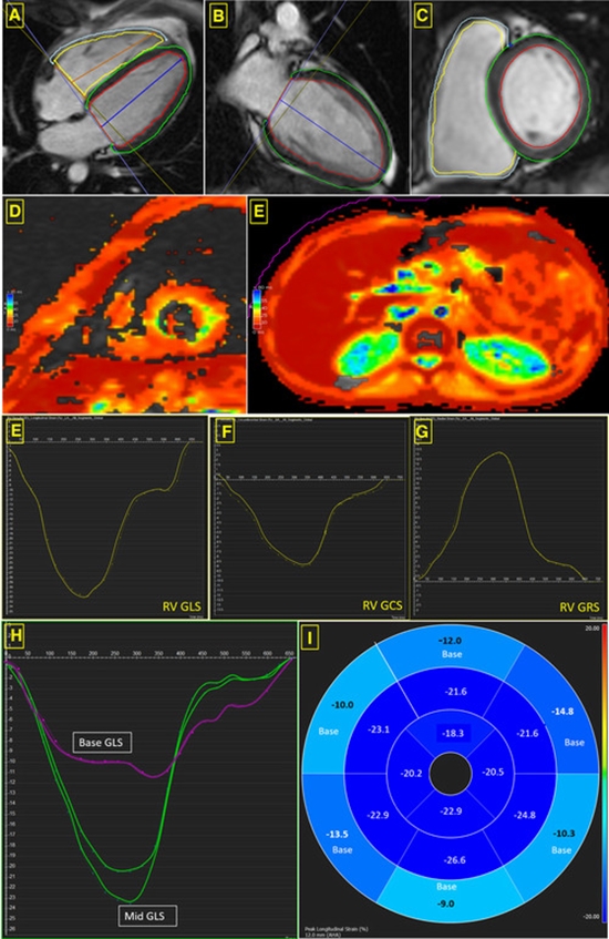
Heart failure resulting from myocardial iron deposition is the most important cause of death in β-thalassemia major (TM) patients. Cardiac T2*magnetic resonance imaging (MRI), echocardiography, and serum ferritin level serve as diagnostic methods for detecting myocardial iron overload. In this study, we aimed to evaluate the relationship between the above-mentioned methods.

Methods

T2*MRI and echocardiographic measurement of left ventricular (LV) systolic and diastolic function were performed in 63 patients. Serum ferritin level was measured. The relationships between all assessments were evaluated.

Results

There were 40 women and 23 men with a mean age of 23.7±5.1 years (range, 15-35 years). There was no statistically significant correlation between serum ferritin level and LV systolic and diastolic function (P=0.994 and P=0.475, respectively). T2*MRI results had a significant correlation with ferritin level; 63.6% of patients with serum ferritin level >2,000 ng/mL had abnormal cardiac MRI, while none of the patients with ferritin level <1,000 ng/mL had abnormal cardiac MRI (P=0.001). There was no significant correlation between MRI findings and LV systolic function (P=1.00). However, we detected a significant difference between LV diastolic function and cardiac siderosis (P=0.03)

Conclusion

MRI findings are a good predictor of future cardiac dysfunction, even in asymptomatic TM patients; however, diastolic dysfunction may happen prior to cardiac siderosis in some patients, and echocardiography is able to diagnose this diastolic dysfunction while T2*MRI shows normal findings.

  • Article_DOI : 10.5045/br.2014.49.3.182
  • Author(s) : farideh moussavi, mounes aliyari ghasabeh
  • News Group : research,research article
  • News Code : 278492
مدیر سایت
Author:

مدیر سایت

Enter your desired term to search
Theme settings