Advanced Diagnostic & Interventional Radiology Research Center | The value of bronchocele attenuation in pulmonary computed tomography in assessment of allergic bronchopulmonary aspergillosis in the background of cystic fibrosis: A cross sectional study

Advanced Diagnostic & Interventional Radiology Research Center | The value of bronchocele attenuation in pulmonary computed tomography in assessment of allergic bronchopulmonary aspergillosis in the background of cystic fibrosis: A cross sectional study
| Dec 11 2025
logo

Advanced Diagnostic & Interventional Radiology Research Center

COVID-19 pandemic 

During the COVID-19 pandemic, the Radiology Research Center at Tehran University of Medical Sciences continued its research activities despite the challenges posed by the increased demand for CT scans of COVID-19 patients and the necessity of adhering to strict health protocols. This center played a crucial role in improving medical imaging techniques, optimizing diagnostic protocols, and advancing technologies related to CT scan image analysis.

Faculty members, researchers, and staff remained committed to ensuring the safety and well-being of healthcare professionals and patients while actively engaging in imaging data analysis, developing artificial intelligence algorithms for faster disease detection, publishing scientific articles, and presenting their findings at international conferences. These efforts aimed to enhance diagnostic accuracy, improve treatment processes, and alleviate pressure on healthcare systems.


Key achievements of the Radiology Research Center during the COVID-19 pandemic include:


✔️ Development and optimization of lung imaging protocols for faster and more accurate COVID-19 diagnosis
✔️ Implementation of artificial intelligence technologies for automated CT scan analysis and reduced diagnosis time
✔️ Publication of high-impact research articles on innovative imaging methods for COVID-19 patients
✔️ Participation in national and international projects focused on COVID-19 diagnosis and patient management

The center remains dedicated to advancing research in medical imaging and continues to contribute as a leading scientific institution in improving the quality of diagnostic and therapeutic services.


Some of the center's significant achievements during the pandemic include:


  • Release Date : Jul 21 2024 - 08:03
  • : 84
  • Study time : 1 minute(s)

The value of bronchocele attenuation in pulmonary computed tomography in assessment of allergic bronchopulmonary aspergillosis in the background of cystic fibrosis: A cross sectional study

 pulmonary computed tomography in assessment of allergic bronchopulmonary {faces}

Background: There is no specific test in the definitive diagnostic approach to Allergic bronchopulmonary aspergillosis (ABPA) especially in the background of cystic fibrosis, but comprehensive and simultaneous clinical, radiological and serological examination will be the basis of ABPA diagnosis. The increasing in attenuation of bronchoceles in imaging has recently been proposed as a valuable diagnostic criterion.

Purpose: The present study aimed to assess bronchocele attenuation in pulmonary CT scan of patients with complicated cystic fibrosis for diagnosis of ABPA.

Methods: This cross-sectional study was performed on 74 consecutive patients aged 3-18 years suffering cystic fibrosis presented with exacerbation of pulmonary symptoms and were suspected of having ABPA. All were examined by 16 Slice CT Scan and the density of bronchoceles above 5 mm in diameter were measured in Hounsfield unit. The total serum IgE titer, skin prick test for aspergillus and anti-aspergillus IgG and IgE level were obtained for all subjects and both cutoff values of IgE level (>500 IU/mL and >1000 IU/mL) were considered as the criteria for ABPA diagnosis.

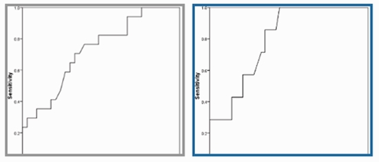
Results: Considering IgE level of greater than 500 IU/mL and 1000 IU/mL as the diagnostic criteria, 24.3% and 10.8% had evidence of ABPA, respectively. Considering the two pointed diagnostic IgE ranges and based on the analysis of the area under the ROC curve, bronchocele attenuation could effectively predict the presence of ABPA with the best cutoff values of 37.25 (with a sensitivity of 70.6% and a specificity of 66.7%) and 40.00 (with a sensitivity of 85.7% and a specificity of 65.1%), respectively.

Conclusion: The presence of bronchocele and an increase in its attenuation on CT scan will be diagnostic for the occurrence of ABPA.

  • Article_DOI : 10.1016/j.amsu.2022.104892
  • Author(s) : mahsa taherzadeh,fatemeh zamani
  • News Group : research,research article
  • News Code : 277953
مدیر سایت
Author:

مدیر سایت

0 Comments for this article

comment

Post your comment:

متن درون تصویر را در جعبه متن زیر وارد نمائید *
Enter your desired term to search
Theme settings