| Jul 23 2025
logo

Advanced Diagnostic & Interventional Radiology Research Center


Artificial Intelligence of Radiology Research Center


Given the vast amount of image data generated daily, radiology research centers possess immense potential for harnessing artificial intelligence. This technology can significantly improve the accuracy and speed of disease diagnosis, aiding radiologists in making informed clinical decisions. Below are some of the activities undertaken by radiology research centers in the realm of AI:


  1. Developing Automated Disease Detection Algorithms:

  • Tumor Detection: Developing algorithms to automatically detect various tumors in radiology images (such as CT scans, MRIs, and PET scans).
  • Pulmonary Disease Detection: Automatically detecting diseases like pneumonia, COVID-19, and pulmonary fibrosis.
  • Cardiovascular Disease Detection: Early detection of cardiovascular diseases such as aortic aneurysm and coronary artery stenosis.
  •  

2. Optimizing Imaging Processes:


  • Reducing Radiation Dose: Developing algorithms to reduce radiation dose during imaging procedures, especially for children and pregnant women.
  • Improving Image Quality: Using AI to enhance radiology image quality and reduce noise.
  • Facilitating Image Interpretation: Developing tools to assist radiologists in interpreting radiology images, such as highlighting suspicious areas.


3. Analyzing Big Data:


  • Identifying Patterns: Utilizing machine learning algorithms to identify hidden patterns in radiology data and discover new correlations between diseases and risk factors.
  • Predicting Disease Progression: Predicting disease progression in patients with chronic conditions.
  • Evaluating Treatment Effectiveness: Assessing the effectiveness of various treatments based on radiology data.
  •  

4. Developing Diagnostic Assistance Tools:


  • Assisting Radiologists: Developing tools to aid radiologists in disease diagnosis, such as automated measurement tools and automatic lesion classification.
  • Interpreting Radiology Reports: Developing systems for automated interpretation of radiology reports and extraction of key information.
  •  

5. Collaborating with Other Fields:


  • Collaborating with Software Engineers: Developing and implementing AI algorithms.
  • Collaborating with Medical Specialists: Validating AI models and ensuring their effectiveness in clinical settings.
  • Collaborating with Data Scientists: Collecting, processing, and analyzing radiology data.


6. Education and Research:


  • Training Personnel: Conducting training courses to familiarize center staff with AI concepts and applications in radiology.
  • Conducting Fundamental Research: Conducting fundamental research in AI for radiology to develop new algorithms and improve existing methods.
  • Publishing Scientific Articles: Publishing research results in reputable scientific journals.
  •  

In conclusion, given the rapid advancements in artificial intelligence, its role in radiology is expected to become increasingly significant in the future. Radiology research centers can make substantial contributions to medical advancements and improved diagnostic services by investing in this field.


Lists of AI articles:


  • Release Date : Sep 23 2024 - 12:44
  • : 84
  • Study time : 1 minute(s)

A novel deep learning model for breast lesion classification using ultrasound Images: A multicenter data evaluation

 {faces}

Purpose: Breast cancer is one of the major reasons of death due to cancer in women. Early diagnosis is the most critical key for disease screening, control, and reducing mortality. A robust diagnosis relies on the correct classification of breast lesions. While breast biopsy is referred to as the "gold standard" in assessing both the activity and degree of breast cancer, it is an invasive and time-consuming approach.

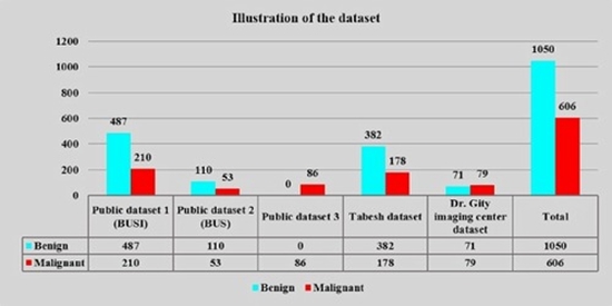
Method: The current study's primary objective was to develop a novel deep-learning architecture based on the InceptionV3 network to classify ultrasound breast lesions. The main promotions of the proposed architecture were converting the InceptionV3 modules to residual inception ones, increasing their number, and altering the hyperparameters. In addition, we used a combination of five datasets (three public datasets and two prepared from different imaging centers) for training and evaluating the model.

Results: The dataset was split into the train (80%) and test (20%) groups. The model achieved 0.83, 0.77, 0.8, 0.81, 0.81, 0.18, and 0.77 for the precision, recall, F1 score, accuracy, AUC, Root Mean Squared Error, and Cronbach's α in the test group, respectively.

Conclusions: This study illustrates that the improved InceptionV3 can robustly classify breast tumors, potentially reducing the need for biopsy in many cases.

Keywords: Breast ultrasound; Convolutional neural network; Deep learning; Image classification.

  • Article_DOI : 10.1016/j.ejmp.2023.102560.
  • Author(s) : nasim sirjani ,mostafa ghelich oghli
  • News Group : research,articles,research article,AI,AI articles
  • News Code : 278846
نفیسه سادات قوامی
Author:

نفیسه سادات قوامی

0 Comments for this article

comment

Post your comment:

متن درون تصویر را در جعبه متن زیر وارد نمائید *
Enter your desired term to search
Theme settings
Close menu