| Jul 23 2025
logo

Advanced Diagnostic & Interventional Radiology Research Center


Artificial Intelligence of Radiology Research Center


Given the vast amount of image data generated daily, radiology research centers possess immense potential for harnessing artificial intelligence. This technology can significantly improve the accuracy and speed of disease diagnosis, aiding radiologists in making informed clinical decisions. Below are some of the activities undertaken by radiology research centers in the realm of AI:


  1. Developing Automated Disease Detection Algorithms:

  • Tumor Detection: Developing algorithms to automatically detect various tumors in radiology images (such as CT scans, MRIs, and PET scans).
  • Pulmonary Disease Detection: Automatically detecting diseases like pneumonia, COVID-19, and pulmonary fibrosis.
  • Cardiovascular Disease Detection: Early detection of cardiovascular diseases such as aortic aneurysm and coronary artery stenosis.
  •  

2. Optimizing Imaging Processes:


  • Reducing Radiation Dose: Developing algorithms to reduce radiation dose during imaging procedures, especially for children and pregnant women.
  • Improving Image Quality: Using AI to enhance radiology image quality and reduce noise.
  • Facilitating Image Interpretation: Developing tools to assist radiologists in interpreting radiology images, such as highlighting suspicious areas.


3. Analyzing Big Data:


  • Identifying Patterns: Utilizing machine learning algorithms to identify hidden patterns in radiology data and discover new correlations between diseases and risk factors.
  • Predicting Disease Progression: Predicting disease progression in patients with chronic conditions.
  • Evaluating Treatment Effectiveness: Assessing the effectiveness of various treatments based on radiology data.
  •  

4. Developing Diagnostic Assistance Tools:


  • Assisting Radiologists: Developing tools to aid radiologists in disease diagnosis, such as automated measurement tools and automatic lesion classification.
  • Interpreting Radiology Reports: Developing systems for automated interpretation of radiology reports and extraction of key information.
  •  

5. Collaborating with Other Fields:


  • Collaborating with Software Engineers: Developing and implementing AI algorithms.
  • Collaborating with Medical Specialists: Validating AI models and ensuring their effectiveness in clinical settings.
  • Collaborating with Data Scientists: Collecting, processing, and analyzing radiology data.


6. Education and Research:


  • Training Personnel: Conducting training courses to familiarize center staff with AI concepts and applications in radiology.
  • Conducting Fundamental Research: Conducting fundamental research in AI for radiology to develop new algorithms and improve existing methods.
  • Publishing Scientific Articles: Publishing research results in reputable scientific journals.
  •  

In conclusion, given the rapid advancements in artificial intelligence, its role in radiology is expected to become increasingly significant in the future. Radiology research centers can make substantial contributions to medical advancements and improved diagnostic services by investing in this field.


Lists of AI articles:


  • Release Date : Sep 23 2024 - 14:23
  • : 92
  • Study time : 1 minute(s)

Risk scores for prediction of paroxysmal atrial fibrillation after acute ischemic stroke or transient ischemic attack: A systematic review and meta-analysis

 {faces}

Introduction: Detection of paroxysmal atrial fibrillation (PAF) is crucial for secondary prevention in patients with recent strokes of unknown etiology. This systematic review and meta-analysis assess the predictive power of available risk scores for detecting new PAF after acute ischemic stroke (AIS).

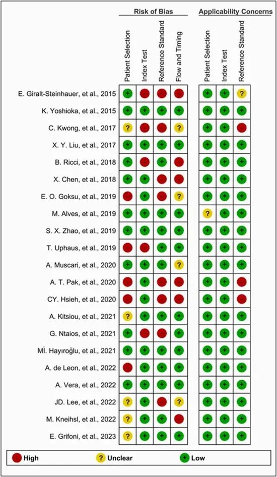
Methods: PubMed, Embase, Scopus, and Web of Science databases were searched until September 2023 to identify relevant studies. A bivariate random effects meta-analysis model pooled data on sensitivity, specificity, and area under the curve (AUC) for each score. The QUADAS-2 tool was used for the quality assessment.

Results: Eventually, 21 studies with 18 original risk scores were identified. Age, left atrial enlargement, and NIHSS score were the most common predictive factors, respectively. Seven risk scores were meta-analyzed, with iPAB showing the highest pooled sensitivity and AUC (sensitivity: 89.4%, specificity: 74.2%, AUC: 0.83), and HAVOC having the highest pooled specificity (sensitivity: 46.3%, specificity: 82.0%, AUC: 0.82). Altogether, seven risk scores displayed good discriminatory power (AUC ≥0.80) with four of them (HAVOC, iPAB, Fujii, and MVP scores) being externally validated.

Conclusion: Available risk scores demonstrate moderate to good predictive accuracy and can help identify patients who would benefit from extended cardiac monitoring after AIS. External validation is essential before widespread clinical adoption.

Keywords: Acute ischemic stroke; Cryptogenic stroke; Paroxysmal atrial fibrillation; Risk score; Risk stratification.


link

  • Article_DOI : 10.1016/j.ijcrp.2024.200249
  • Author(s) : sina kazemian,diana zarei , ali bozorgi,saman nazarian ,mahbod issaiy
  • News Group : research,articles,research article,AI,AI articles
  • News Code : 278862
نفیسه سادات قوامی
Author:

نفیسه سادات قوامی

0 Comments for this article

comment

Post your comment:

متن درون تصویر را در جعبه متن زیر وارد نمائید *
Enter your desired term to search
Theme settings
Close menu