Advanced Diagnostic & Interventional Radiology Research Center | A Spectrum of Ultrasound and MR Imaging of Fetal Gastrointestinal Abnormalities. Part 1: Esophagus to Colon

Advanced Diagnostic & Interventional Radiology Research Center | A Spectrum of Ultrasound and MR Imaging of Fetal Gastrointestinal Abnormalities. Part 1: Esophagus to Colon
| Dec 13 2025
logo

Advanced Diagnostic & Interventional Radiology Research Center

COVID-19 pandemic 

During the COVID-19 pandemic, the Radiology Research Center at Tehran University of Medical Sciences continued its research activities despite the challenges posed by the increased demand for CT scans of COVID-19 patients and the necessity of adhering to strict health protocols. This center played a crucial role in improving medical imaging techniques, optimizing diagnostic protocols, and advancing technologies related to CT scan image analysis.

Faculty members, researchers, and staff remained committed to ensuring the safety and well-being of healthcare professionals and patients while actively engaging in imaging data analysis, developing artificial intelligence algorithms for faster disease detection, publishing scientific articles, and presenting their findings at international conferences. These efforts aimed to enhance diagnostic accuracy, improve treatment processes, and alleviate pressure on healthcare systems.


Key achievements of the Radiology Research Center during the COVID-19 pandemic include:


✔️ Development and optimization of lung imaging protocols for faster and more accurate COVID-19 diagnosis
✔️ Implementation of artificial intelligence technologies for automated CT scan analysis and reduced diagnosis time
✔️ Publication of high-impact research articles on innovative imaging methods for COVID-19 patients
✔️ Participation in national and international projects focused on COVID-19 diagnosis and patient management

The center remains dedicated to advancing research in medical imaging and continues to contribute as a leading scientific institution in improving the quality of diagnostic and therapeutic services.


Some of the center's significant achievements during the pandemic include:


  • Release Date : Jul 14 2024 - 07:29
  • : 118
  • Study time : Less than one minute

A Spectrum of Ultrasound and MR Imaging of Fetal Gastrointestinal Abnormalities. Part 1: Esophagus to Colon

Ultrasound and MR Imaging of Fetal Gastrointestinal Abnormalities {faces}

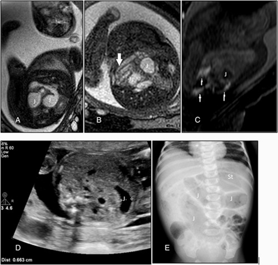
Ultrasound (US) and magnetic resonance imaging (MRI) are two modalities for diagnosing fetal gastrointestinal (GI) anomalies. Ultrasound (US) is the modality of choice. MRI can be used as a complementary method. Despite its expanding utilization in central nervous system (CNS) fetal malformation, MRI has not yet been established for evaluation of fetal GI abnormalities. Therefore, more attention should be paid to the clinical implications of MRI investigations following screening by US

  • Article_DOI : 10.1002/jum.15932
  • Author(s) : behnaz moradi ,ahmad-reza tahmasebpour,masoumeh banihashemian,amir reza radmard
  • News Group : research,research article
  • News Code : 277974
مدیر سایت
Author:

مدیر سایت

Enter your desired term to search
Theme settings