Advanced Diagnostic & Interventional Radiology Research Center | biomarker quantification methodology for DCE-MRI of adnexal mass

Advanced Diagnostic & Interventional Radiology Research Center | biomarker quantification methodology for DCE-MRI of adnexal mass
| Dec 12 2025
logo

Advanced Diagnostic & Interventional Radiology Research Center

COVID-19 pandemic 

During the COVID-19 pandemic, the Radiology Research Center at Tehran University of Medical Sciences continued its research activities despite the challenges posed by the increased demand for CT scans of COVID-19 patients and the necessity of adhering to strict health protocols. This center played a crucial role in improving medical imaging techniques, optimizing diagnostic protocols, and advancing technologies related to CT scan image analysis.

Faculty members, researchers, and staff remained committed to ensuring the safety and well-being of healthcare professionals and patients while actively engaging in imaging data analysis, developing artificial intelligence algorithms for faster disease detection, publishing scientific articles, and presenting their findings at international conferences. These efforts aimed to enhance diagnostic accuracy, improve treatment processes, and alleviate pressure on healthcare systems.


Key achievements of the Radiology Research Center during the COVID-19 pandemic include:


✔️ Development and optimization of lung imaging protocols for faster and more accurate COVID-19 diagnosis
✔️ Implementation of artificial intelligence technologies for automated CT scan analysis and reduced diagnosis time
✔️ Publication of high-impact research articles on innovative imaging methods for COVID-19 patients
✔️ Participation in national and international projects focused on COVID-19 diagnosis and patient management

The center remains dedicated to advancing research in medical imaging and continues to contribute as a leading scientific institution in improving the quality of diagnostic and therapeutic services.


Some of the center's significant achievements during the pandemic include:


  • Release Date : Apr 7 2024 - 12:40
  • : 8
  • Study time : 1 minute(s)

A one-step biomarker quantification methodology for DCE-MRI of adnexal masses: Capturing kinetic pattern from early to late enhancement

biomarker quantification methodology for DCE-MRI of adnexal masses {faces}

Purpose: To develop a one-step quantification approach that accounts for joint preprocessing and quantification of whole-range kinetics (early and late-phase washout) of dynamic contrast-enhanced (DCE) MRI of indeterminate adnexal masses.

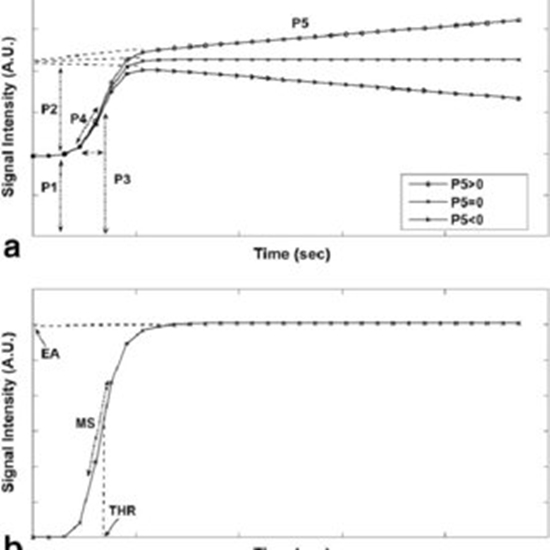
Methods: Preoperative DCE-MRI of 43 (24 benign, 19 malignant) sonographically indeterminate adnexal masses were analyzed prospectively. A five-parameter sigmoid function was implemented to model the enhancement curves calculated within regions of interest. Diagnostic performance of five-parameter sigmoid model parameters (P1 through P5 ) was compared with pharmacokinetic (PK) modeling, semiquantitative analysis, and three-parameter sigmoid. Statistical analysis was performed using two-tailed student's t-test.

Results: The results revealed that P2 , representing the enhancement amplitude, is significantly higher, and P5 , indicating the terminal phase, is generally negative in malignant lesions (P < 0.001). P2 (sensitivity = 79%, specificity = 87.5%, accuracy = 84%, area under the receiver operating characteristic curve = 91%) outperforms classification performances of PK and semiquantitative parameters. A combination of P2 and P5 shows comparable performance (sensitivity = 79%, specificity = 87.5%, accuracy = 84%, area under the receiver operating characteristic curve = 92%) to that of the combination of PK parameters, whereas the five-parameter sigmoid function maintains fewer assumptions than PK.

Conclusions: The presented one-step quantification approach is helpful for accurate discrimination of benign from malignant indeterminate adnexal masses. Accordingly, P2 has considerably high diagnostic performance and terminal slope (P5 ), as a previously overlooked feature, contributes more than widely accepted early-enhancement kinetic features. Magn Reson Med 79:1165-1171, 2018. © 2017 International Society for Magnetic Resonance in Medicine

  • Article_DOI : 10.1002/mrm.26743
  • Author(s) : hamidreza saligheh rad,anahita fathi kazerooni
  • News Group : research,research article
  • News Code : 278389
مدیر سایت
Author:

مدیر سایت

Enter your desired term to search
Theme settings