Advanced Diagnostic & Interventional Radiology Research Center | Accurate quantification of choline to creatine ratio as a biomarker to distinguish osteosarcoma patients from normal subjects employing proton magnetic resonance spectroscopy imaging at 3 tesla

Advanced Diagnostic & Interventional Radiology Research Center | Accurate quantification of choline to creatine ratio as a biomarker to distinguish osteosarcoma patients from normal subjects employing proton magnetic resonance spectroscopy imaging at 3 tesla
| Dec 10 2025
logo

Advanced Diagnostic & Interventional Radiology Research Center

COVID-19 pandemic 

During the COVID-19 pandemic, the Radiology Research Center at Tehran University of Medical Sciences continued its research activities despite the challenges posed by the increased demand for CT scans of COVID-19 patients and the necessity of adhering to strict health protocols. This center played a crucial role in improving medical imaging techniques, optimizing diagnostic protocols, and advancing technologies related to CT scan image analysis.

Faculty members, researchers, and staff remained committed to ensuring the safety and well-being of healthcare professionals and patients while actively engaging in imaging data analysis, developing artificial intelligence algorithms for faster disease detection, publishing scientific articles, and presenting their findings at international conferences. These efforts aimed to enhance diagnostic accuracy, improve treatment processes, and alleviate pressure on healthcare systems.


Key achievements of the Radiology Research Center during the COVID-19 pandemic include:


✔️ Development and optimization of lung imaging protocols for faster and more accurate COVID-19 diagnosis
✔️ Implementation of artificial intelligence technologies for automated CT scan analysis and reduced diagnosis time
✔️ Publication of high-impact research articles on innovative imaging methods for COVID-19 patients
✔️ Participation in national and international projects focused on COVID-19 diagnosis and patient management

The center remains dedicated to advancing research in medical imaging and continues to contribute as a leading scientific institution in improving the quality of diagnostic and therapeutic services.


Some of the center's significant achievements during the pandemic include:


  • Release Date : Jul 10 2024 - 10:45
  • : 81
  • Study time : 1 minute(s)

Accurate quantification of choline to creatine ratio as a biomarker to distinguish osteosarcoma patients from normal subjects employing proton magnetic resonance spectroscopy imaging at 3 tesla

choline-to-creatine ratio as a biomarker to distinguish osteosarcoma patients {faces}

Purpose: This study focused on accurate quantification of a maximum of Choline-to-Creatine ratio (Max (Cho/Cr)) in 10 Osteosarcoma patients, in comparison with 5 healthy volunteers as our control group using proton Magnetic Resonance Spectroscopy Imaging (1H-MRSI).

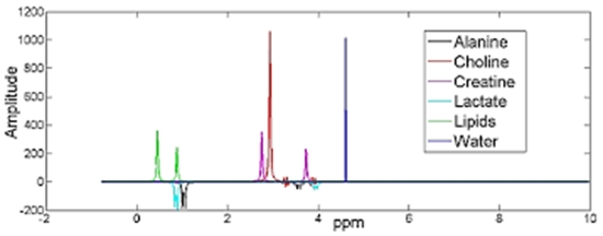
Materials and Methods: Max (Cho/Cr) were obtained in 10 patients with Osteosarcoma over their corresponding ratio maps containing diseased tissue, to be compared with Cho/Cr in 5 healthy volunteers at 3T, employing MRSI (Performed Employing Pointed-resolved Spectroscopy (PRESS), TR/TE: 2500s /135 ms) with water-suppression. An extra unsuppressed water Single-Voxel Spectroscopy (SVS) was acquired to provide phase information for further Eddy Current Correction (ECC). Multi-stage preprocessing was applied. Subtract QUEST MRSI as a time-domain technique was employed to accurately quantify the metabolites’ ratios and to estimate the baseline.

Results: An optimal database for Subtract QUEST was achieved based on multiple trials evaluated by acceptable peak-fitting and Cramer-Rao-Bound (CRB). Lipids at frequencies of 0.94 and 1.33ppm were combined to increase the accuracy of the Lipid estimation.

Conclusion: Estimation of Max (Cho/Cr) evaluated over Cho/Cr spatial maps to distinguish Osteosarcoma patients from normal subjects suggested that the proposed quantification method leads to high power and linear classifier with a high degree of reproducibility, considering 1H-MRSI at 3T machine as a high efficacy diagnostic tool for musculoskeletal radiology.

  • Article_DOI : https://doi.org/10.18502/fbt.v8i2.6513
  • Author(s) : shaghayegh karimi alavijeh,fakhereh pashaei,mahrooz malek,hamidreza saligheh rad
  • News Group : research,research article
  • News Code : 277990
مدیر سایت
Author:

مدیر سایت

Enter your desired term to search
Theme settings