Advanced Diagnostic & Interventional Radiology Research Center | Application of ultrasound elastography for determining carpal tu

Advanced Diagnostic & Interventional Radiology Research Center | Application of ultrasound elastography for determining carpal tu
| Jan 2 2026
logo

Advanced Diagnostic & Interventional Radiology Research Center

COVID-19 pandemic 

During the COVID-19 pandemic, the Radiology Research Center at Tehran University of Medical Sciences continued its research activities despite the challenges posed by the increased demand for CT scans of COVID-19 patients and the necessity of adhering to strict health protocols. This center played a crucial role in improving medical imaging techniques, optimizing diagnostic protocols, and advancing technologies related to CT scan image analysis.

Faculty members, researchers, and staff remained committed to ensuring the safety and well-being of healthcare professionals and patients while actively engaging in imaging data analysis, developing artificial intelligence algorithms for faster disease detection, publishing scientific articles, and presenting their findings at international conferences. These efforts aimed to enhance diagnostic accuracy, improve treatment processes, and alleviate pressure on healthcare systems.


Key achievements of the Radiology Research Center during the COVID-19 pandemic include:


✔️ Development and optimization of lung imaging protocols for faster and more accurate COVID-19 diagnosis
✔️ Implementation of artificial intelligence technologies for automated CT scan analysis and reduced diagnosis time
✔️ Publication of high-impact research articles on innovative imaging methods for COVID-19 patients
✔️ Participation in national and international projects focused on COVID-19 diagnosis and patient management

The center remains dedicated to advancing research in medical imaging and continues to contribute as a leading scientific institution in improving the quality of diagnostic and therapeutic services.


Some of the center's significant achievements during the pandemic include:


  • Release Date : Mar 3 2024 - 12:59
  • : 43
  • Study time : 1 minute(s)

Application of ultrasound elastography for determining carpal tunnel syndrome severity

To evaluate grey-scale and elastography ultrasound imaging findings in patients with CTS compared to nerve conductive studies.

ultrasound elastography for determining carpal tunnel syndrome {faces}

Objective: To evaluate grey-scale and elastography ultrasound imaging findings in patients with CTS compared to nerve conductive studies.

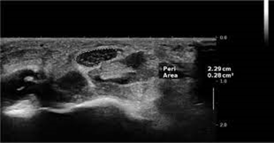
Method: Sixty median nerves of 31 cases with confirmed CTS and 44 median nerves in 22 controls (healthy volunteers) who had no clinical evidence of CTS were evaluated. An expert radiologist performed all US evaluations. The RGB image is a three-dimensional matrix. A colour image RGB is an M × N × 3 array of colour pixels. The total pixels, total blue and red pixels, and blue and red indexes were compared between cases and controls.

Results: Of the 60 nerves in the cases, 17 (16.3%) were mildly affected, 30 (28.8%) were moderately affected, and 13 (12.5%) were severely affected. Mean CSA, total blue pixels and blue indexes were significantly different between controls and cases with different levels of disease severity. The best cut-off point in the blue index to differentiate patients from controls was 0.1486, with a sensitivity and specificity of 80 and 70% (AUC = 0.79, P < 0.001), respectively. The best cut-off point for the red index to differentiate patients from controls was 0.1896, with a sensitivity and specificity of 70 and 55% (AUC = 0.64, P = 0.01), respectively.

Conclusion: Sono-elastography could be a useful diagnostic method for evaluating CTS severity in affected cases.

  • Article_DOI : 10.1007/s11604-015-0416-3
  • Author(s) : mahrooz malek,mahsa ghajarzadeh
  • News Group : research article,AI,research
  • News Code : 278467
مدیر سایت
Author:

مدیر سایت

Enter your desired term to search
Theme settings