Advanced Diagnostic & Interventional Radiology Research Center | contrast of the calcification in digital mammography system

Advanced Diagnostic & Interventional Radiology Research Center | contrast of the calcification in digital mammography system
| Dec 12 2025
logo

Advanced Diagnostic & Interventional Radiology Research Center

COVID-19 pandemic 

During the COVID-19 pandemic, the Radiology Research Center at Tehran University of Medical Sciences continued its research activities despite the challenges posed by the increased demand for CT scans of COVID-19 patients and the necessity of adhering to strict health protocols. This center played a crucial role in improving medical imaging techniques, optimizing diagnostic protocols, and advancing technologies related to CT scan image analysis.

Faculty members, researchers, and staff remained committed to ensuring the safety and well-being of healthcare professionals and patients while actively engaging in imaging data analysis, developing artificial intelligence algorithms for faster disease detection, publishing scientific articles, and presenting their findings at international conferences. These efforts aimed to enhance diagnostic accuracy, improve treatment processes, and alleviate pressure on healthcare systems.


Key achievements of the Radiology Research Center during the COVID-19 pandemic include:


✔️ Development and optimization of lung imaging protocols for faster and more accurate COVID-19 diagnosis
✔️ Implementation of artificial intelligence technologies for automated CT scan analysis and reduced diagnosis time
✔️ Publication of high-impact research articles on innovative imaging methods for COVID-19 patients
✔️ Participation in national and international projects focused on COVID-19 diagnosis and patient management

The center remains dedicated to advancing research in medical imaging and continues to contribute as a leading scientific institution in improving the quality of diagnostic and therapeutic services.


Some of the center's significant achievements during the pandemic include:


  • Release Date : Apr 6 2024 - 11:30
  • : 29
  • Study time : 1 minute(s)

Calculation of the contrast of the calcification in digital mammography system: Gate validation

Calculation of the contrast of the calcification in digital mammography {faces}

Purpose: Validation of the Gate tool in digital mammography image simulation from the viewpoint of image quality (contrast of calcifications).

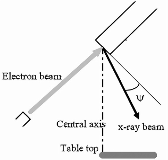
Materials and methods: The polymethyl methacrylate (PMMA) phantom containing aluminum foils in different thicknesses is used for measuring the contrast of calcifications in a real system. In this research, the phantom and mammography system have been simulated by the Gate tool with the maximum possible details. The contrast of the aluminum foil in simulations and practical method has been compared with each other and the standard errors in the mean (SEM) for various voltages of X-ray tube, aluminum foil, and PMMA thicknesses have been reported.

Results: Based on the obtained results, by increasing the X-ray tube voltage from 20 to 39 kVp, the image contrast has been decreased in both simulation and practical methods. The minimum and maximum average SEM of the contrast of the aluminum foils among various voltages between two simulations and practical methods for different PMMA thicknesses of 2, 4, and 6 cm have been reported as 0.0105 and 0.0117, 0.0049 and 0.0154, and 0.0037 and 0.0072, respectively.

Discussion: According to the SEM rate reported in this research for calculating the contrast of the aluminum foils in the mammography system based on simulation and practical methods, the capability of the Gate tool for simulating digital mammography system and the images created in it from the viewpoint of image contrast can be confirmed.

  • Article_DOI : 10.4103/0973-1482.168967
  • Author(s) : dooman arefan,alireza talebpour
  • News Group : research,research article
  • News Code : 278393
مدیر سایت
Author:

مدیر سایت

Enter your desired term to search
Theme settings