Advanced Diagnostic & Interventional Radiology Research Center | Cardiac magnetic resonance imaging in myocardial infarction

Advanced Diagnostic & Interventional Radiology Research Center | Cardiac magnetic resonance imaging in myocardial infarction
| Dec 10 2025
logo

Advanced Diagnostic & Interventional Radiology Research Center

COVID-19 pandemic 

During the COVID-19 pandemic, the Radiology Research Center at Tehran University of Medical Sciences continued its research activities despite the challenges posed by the increased demand for CT scans of COVID-19 patients and the necessity of adhering to strict health protocols. This center played a crucial role in improving medical imaging techniques, optimizing diagnostic protocols, and advancing technologies related to CT scan image analysis.

Faculty members, researchers, and staff remained committed to ensuring the safety and well-being of healthcare professionals and patients while actively engaging in imaging data analysis, developing artificial intelligence algorithms for faster disease detection, publishing scientific articles, and presenting their findings at international conferences. These efforts aimed to enhance diagnostic accuracy, improve treatment processes, and alleviate pressure on healthcare systems.


Key achievements of the Radiology Research Center during the COVID-19 pandemic include:


✔️ Development and optimization of lung imaging protocols for faster and more accurate COVID-19 diagnosis
✔️ Implementation of artificial intelligence technologies for automated CT scan analysis and reduced diagnosis time
✔️ Publication of high-impact research articles on innovative imaging methods for COVID-19 patients
✔️ Participation in national and international projects focused on COVID-19 diagnosis and patient management

The center remains dedicated to advancing research in medical imaging and continues to contribute as a leading scientific institution in improving the quality of diagnostic and therapeutic services.


Some of the center's significant achievements during the pandemic include:


  • Release Date : Jul 15 2025 - 07:01
  • : 37
  • Study time : 1 minute(s)

Cardiac magnetic resonance imaging in myocardial infarction with non obstructed coronary arteries: diagnostic and prognostic value

 {faces}

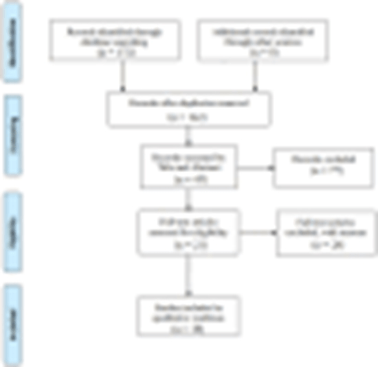
Myocardial infarction with non-obstructed coronary arteries (MINOCA) occurs when patients experience a heart attack without significant coronary artery blockage despite showing acute coronary syndrome symptoms. Unlike stable atherosclerosis, MINOCA involves acute myocardial infarction (MI) without obstructive coronary artery disease (CAD). The diagnostic criteria included meeting the universal MI definition, non-obstructive coronary arteries on angiography (< 50% stenosis), and no apparent cause of the acute event. The causes include coronary, cardiac, and extracardiac origins, such as plaque rupture, coronary spasm, myocarditis, or pulmonary embolism. MINOCA affects 5-6% of patients with acute MI undergoing angiography, with variations based on demographic factors. Although MINOCA was initially believed to have a favorable outcome, recent findings have indicated that MINOCA patients have a worse prognosis than the general population. Current guidelines strongly advocate the use of cardiac magnetic resonance imaging (CMR) to evaluate suspected MINOCA cases. However, multiple studies have demonstrated that CMR may fail to detect some instances of MINOCA, particularly in cases of mild inflammation or minor infractions. This could lead to a false-negative diagnosis requiring further testing. This review aimed to evaluate the diagnostic and prognostic value of CMR in patients with potential MINOCA.

  • Article_DOI : 10.62347/FCDC4114
  • Author(s) : farshad riahi
  • News Group : research,research article
  • News Code : 300919
نفیسه سادات قوامی
Author:

نفیسه سادات قوامی

0 Comments for this article

comment

Post your comment:

متن درون تصویر را در جعبه متن زیر وارد نمائید *
Enter your desired term to search
Theme settings