Advanced Diagnostic & Interventional Radiology Research Center | MRI in Detecting Axillary Lymph Node Metastasis in Breast Cancer

Advanced Diagnostic & Interventional Radiology Research Center | MRI in Detecting Axillary Lymph Node Metastasis in Breast Cancer
| Aug 3 2026
logo

Advanced Diagnostic & Interventional Radiology Research Center

COVID-19 pandemic 

During the COVID-19 pandemic, the Radiology Research Center at Tehran University of Medical Sciences continued its research activities despite the challenges posed by the increased demand for CT scans of COVID-19 patients and the necessity of adhering to strict health protocols. This center played a crucial role in improving medical imaging techniques, optimizing diagnostic protocols, and advancing technologies related to CT scan image analysis.

Faculty members, researchers, and staff remained committed to ensuring the safety and well-being of healthcare professionals and patients while actively engaging in imaging data analysis, developing artificial intelligence algorithms for faster disease detection, publishing scientific articles, and presenting their findings at international conferences. These efforts aimed to enhance diagnostic accuracy, improve treatment processes, and alleviate pressure on healthcare systems.


Key achievements of the Radiology Research Center during the COVID-19 pandemic include:


✔️ Development and optimization of lung imaging protocols for faster and more accurate COVID-19 diagnosis
✔️ Implementation of artificial intelligence technologies for automated CT scan analysis and reduced diagnosis time
✔️ Publication of high-impact research articles on innovative imaging methods for COVID-19 patients
✔️ Participation in national and international projects focused on COVID-19 diagnosis and patient management

The center remains dedicated to advancing research in medical imaging and continues to contribute as a leading scientific institution in improving the quality of diagnostic and therapeutic services.


Some of the center's significant achievements during the pandemic include:


  • Release Date : Jun 16 2026 - 10:13
  • : 2
  • Study time : 1 minute(s)

Comparative Analysis of Abbreviated and Full Protocol MRI in Detecting Axillary Lymph Node Metastasis in Patients with Known Breast Cancer
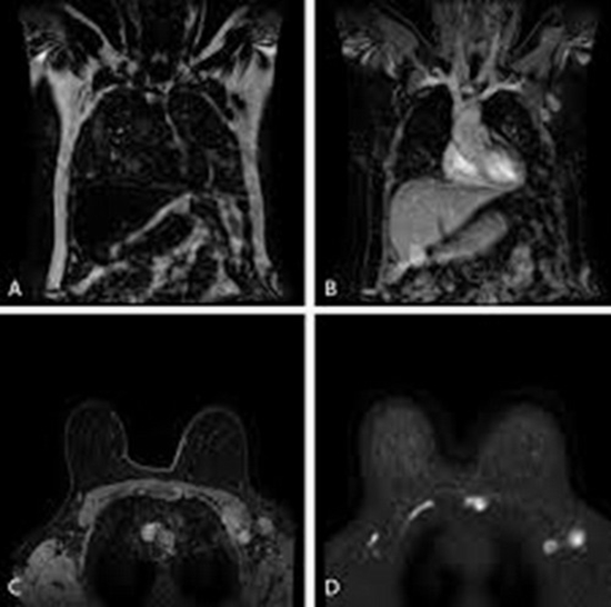
MRI in Detecting Axillary Lymph Node Metastasis in Breast Cancer {faces}

Background: Accurate axillary lymph node staging is crucial for breast cancer prognosis and treatment planning. This study compares the diagnostic efficacy of abbreviated MRI (AB-MRI) protocols with limited sequences and reduced time, against full-diagnostic MRI (FD-MRI) in staging axillary lymph node metastasis of breast cancer patients.

Methods: This was a retrospective cross-sectional diagnostic-accuracy study of 88 women with breast cancer who underwent MRI for axillary lymph node staging. MRI protocols included FD-MRI, non-contrast T1 sequence, and contrast-enhanced T1 sequence. Imaging findings, interpreted by two radiologists blinded to histopathological results, were correlated with findings from sentinel lymph node biopsy or axillary lymph node dissection as the gold standard. Data analysis comprised diagnostic performance parameters (sensitivity and specificity) and inter-protocol agreement using the kappa statistic.

Results: No statistically significant differences were detected among the three protocols (all McNemar's p-values > 0.05). The non-contrast abbreviated MRI protocol demonstrated a sensitivity of 84.9% (95% CI: 72.4%-93.3%) and a specificity of 85.7% (95% CI: 69.7%-95.2%). Unweighted Cohen’s Kappa demonstrated strong concordance between the non-contrast and contrast-enhanced AB-MRI protocols (κ = 0.931; 95% CI: 0.855–1.00), between the non-contrast AB-MRI protocol and the FD-MRI (κ = 0.930; 95% CI: 0.852–1.00), and between the contrast-enhanced AB-MRI protocol and the FD-MRI (κ = 0.907; 95% CI: 0.819–0.995), respectively.

Conclusion: Non-contrast AB-MRI provides a less invasive, cost-effective alternative to FD-MRI for staging axillary lymph nodes in breast cancer, with shorter scan times and fewer procedural risks. Further studies are needed for validation in larger cohorts.

  • Article_DOI : doi.org/10.32768/abc.9268135740-268
  • Author(s) : fahimeh zeinalkhani,mahdieh movahedi
  • News Group : research,research article
  • News Code : 323629
نفیسه سادات قوامی
Author:

نفیسه سادات قوامی

0 Comments for this article

comment

Post your comment:

متن درون تصویر را در جعبه متن زیر وارد نمائید *
Enter your desired term to search
Theme settings