Advanced Diagnostic & Interventional Radiology Research Center | Comparison of Gadovist and Magnevist in Brain mri of MS

Advanced Diagnostic & Interventional Radiology Research Center | Comparison of Gadovist and Magnevist in Brain mri of MS
| Dec 10 2025
logo

Advanced Diagnostic & Interventional Radiology Research Center

COVID-19 pandemic 

During the COVID-19 pandemic, the Radiology Research Center at Tehran University of Medical Sciences continued its research activities despite the challenges posed by the increased demand for CT scans of COVID-19 patients and the necessity of adhering to strict health protocols. This center played a crucial role in improving medical imaging techniques, optimizing diagnostic protocols, and advancing technologies related to CT scan image analysis.

Faculty members, researchers, and staff remained committed to ensuring the safety and well-being of healthcare professionals and patients while actively engaging in imaging data analysis, developing artificial intelligence algorithms for faster disease detection, publishing scientific articles, and presenting their findings at international conferences. These efforts aimed to enhance diagnostic accuracy, improve treatment processes, and alleviate pressure on healthcare systems.


Key achievements of the Radiology Research Center during the COVID-19 pandemic include:


✔️ Development and optimization of lung imaging protocols for faster and more accurate COVID-19 diagnosis
✔️ Implementation of artificial intelligence technologies for automated CT scan analysis and reduced diagnosis time
✔️ Publication of high-impact research articles on innovative imaging methods for COVID-19 patients
✔️ Participation in national and international projects focused on COVID-19 diagnosis and patient management

The center remains dedicated to advancing research in medical imaging and continues to contribute as a leading scientific institution in improving the quality of diagnostic and therapeutic services.


Some of the center's significant achievements during the pandemic include:


  • Release Date : Jul 10 2024 - 11:57
  • : 103
  • Study time : 1 minute(s)

Comparison of Gadovist and Magnevist in Brain Magnetic Resonance Imaging of Multiple Sclerosis Patients with an Acute Attack

Comparison of Gadovist and Magnevist in Brain Magnetic Resonance Imaging of Multiple Sclerosis  {faces}

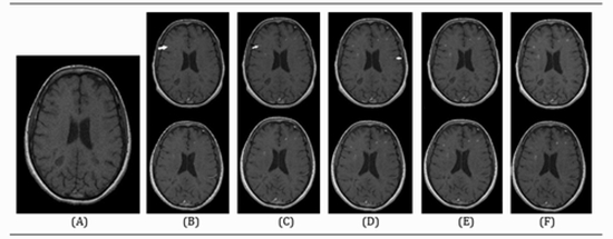
 Background: The use of an appropriate contrast agent performs a major role in brain Magnetic resonance imaging (MRI) of Multiple sclerosis (MS) patients. Objectives: The present study aimed to make a comparison between the diagnostic values of Gadovist and Magnevist considering the successive imaging times in contrast-enhanced brain MRI of MS patients. Methods: A total of 62 relapsing-remitting MS patients (56 females, mean age of 31 years) were enrolled in the present study. All of them underwent two sessions of standard contrast-enhanced brain MRI upon enrollment and 48 h later. The participants were randomly assigned to each contrast agent. T1-weighted (T1W) images were taken 30 sec, as well as 5, 10, 15, and 30 min after the contrast injection. For all of the images, two neuro-radiologists who were blinded to the contrast type counted the number of plaques in the brain. In addition, for the enhanced plaques larger than 10 mm, the signal intensity (SI) was determined using its region of interest. Results: The mean plaque number significantly increased from 30 sec to 15 min for both contrasts separately (P<0. 001). Nonetheless, the slight increases in the mean plaque number from 15-30 min for both Gadovist and Magnevist were not statistically significant (both PValues>0. 25). The mean plaque number in the Gadovist group was higher, compared to that in the Magnevist group at both 15 and 30 min, and the differences were statistically on the borderline (both P-Values=0. 07). The mean SI of enhanced plaques gradually increased in the course of imaging in both contrast groups. Except for 30 sec, in all other time sessions, the mean SI was higher in Gadovistenhanced MR images, compared to Magnevist-enhanced MR images (P<0. 01). Conclusion: As evidenced by the obtained results, Gadovist showed a relatively better diagnostic value for brain MRI of MS patients. Furthermore, the findings suggested that it is cost-effective to take MRI only up to 15 min (instead of 30 min) after contrast injection in both agents

  • Article_DOI : 10.32592/ircmj.2021.23.2.192
  • Author(s) : hossein ghanaati,hassan hashemi
  • News Group : research,research article
  • News Code : 277985
مدیر سایت
Author:

مدیر سایت

Enter your desired term to search
Theme settings