Advanced Diagnostic & Interventional Radiology Research Center | diffusion and peritumoral edema with pathologic in breast cance

Advanced Diagnostic & Interventional Radiology Research Center | diffusion and peritumoral edema with pathologic in breast cance
| Feb 3 2026
logo

Advanced Diagnostic & Interventional Radiology Research Center

COVID-19 pandemic 

During the COVID-19 pandemic, the Radiology Research Center at Tehran University of Medical Sciences continued its research activities despite the challenges posed by the increased demand for CT scans of COVID-19 patients and the necessity of adhering to strict health protocols. This center played a crucial role in improving medical imaging techniques, optimizing diagnostic protocols, and advancing technologies related to CT scan image analysis.

Faculty members, researchers, and staff remained committed to ensuring the safety and well-being of healthcare professionals and patients while actively engaging in imaging data analysis, developing artificial intelligence algorithms for faster disease detection, publishing scientific articles, and presenting their findings at international conferences. These efforts aimed to enhance diagnostic accuracy, improve treatment processes, and alleviate pressure on healthcare systems.


Key achievements of the Radiology Research Center during the COVID-19 pandemic include:


✔️ Development and optimization of lung imaging protocols for faster and more accurate COVID-19 diagnosis
✔️ Implementation of artificial intelligence technologies for automated CT scan analysis and reduced diagnosis time
✔️ Publication of high-impact research articles on innovative imaging methods for COVID-19 patients
✔️ Participation in national and international projects focused on COVID-19 diagnosis and patient management

The center remains dedicated to advancing research in medical imaging and continues to contribute as a leading scientific institution in improving the quality of diagnostic and therapeutic services.


Some of the center's significant achievements during the pandemic include:


  • Release Date : Jun 19 2024 - 08:24
  • : 54
  • Study time : 1 minute(s)

Correlation of apparent diffusion coefficient values and peritumoral edema with pathologic biomarkers in patients with breast cancer

peritumoral edema with pathologic biomarkers in patients with breast cancer {faces}

Purpose: To investigate the relationship between breast cancer imaging features on magnetic resonance imaging (MRI) and histopathological characteristics.

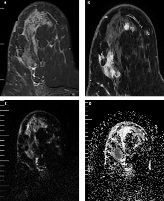
Methods and materials: We prospectively enrolled 46 patients who underwent 1.5-T MRI with 68 breast malignant lesions from 2017 until 2019. Peritumoral edema was determined based on visual assessment on T2 weighted imaging. Lesions were categorized into two groups: A: with edema (48 lesions) and B: without edema (20 lesions).

Results: The tumor size was not different among two groups but multifocal-multicentric lesions were more common in the group A (70% vs. 35%). The axillary lymph nodes are most involved in group A. ER and PR positive lesions were more common in group B (90% vs. 56.3%) but in the group A, HER2 positive lesions were found to be more common (31.3% vs. 15%). The mean ADC value in tumors and peritumoral regions were lower (0.97 × 10⁻³ mm2/s, P = 0.023) and higher (1.85 × 10⁻³ mm2/s, P < 0.0001) in group A, respectively. Peritumoral ADC value was significantly higher in HER2-positive group.

Conclusion: Breast carcinomas with peritumoral edema were found to be more multifocal-multicentric, with higher prevalence of axillary lymph node involvement, more HER 2-positive, with lower prevalence of ER/PR-positive, lower tumoral ADC and higher peritumoral ADC values


  • Article_DOI : 10.1016/j.clinimag.2020.08.020
  • Author(s) : behnaz moradi ,mohammad ali kazemi
  • News Group : research,research article
  • News Code : 278286
مدیر سایت
Author:

مدیر سایت

Enter your desired term to search
Theme settings