Advanced Diagnostic & Interventional Radiology Research Center | Diagnostic Performance of Sonoelastography in Addition to Ultras

Advanced Diagnostic & Interventional Radiology Research Center | Diagnostic Performance of Sonoelastography in Addition to Ultras
| Jan 1 2026
logo

Advanced Diagnostic & Interventional Radiology Research Center

COVID-19 pandemic 

During the COVID-19 pandemic, the Radiology Research Center at Tehran University of Medical Sciences continued its research activities despite the challenges posed by the increased demand for CT scans of COVID-19 patients and the necessity of adhering to strict health protocols. This center played a crucial role in improving medical imaging techniques, optimizing diagnostic protocols, and advancing technologies related to CT scan image analysis.

Faculty members, researchers, and staff remained committed to ensuring the safety and well-being of healthcare professionals and patients while actively engaging in imaging data analysis, developing artificial intelligence algorithms for faster disease detection, publishing scientific articles, and presenting their findings at international conferences. These efforts aimed to enhance diagnostic accuracy, improve treatment processes, and alleviate pressure on healthcare systems.


Key achievements of the Radiology Research Center during the COVID-19 pandemic include:


✔️ Development and optimization of lung imaging protocols for faster and more accurate COVID-19 diagnosis
✔️ Implementation of artificial intelligence technologies for automated CT scan analysis and reduced diagnosis time
✔️ Publication of high-impact research articles on innovative imaging methods for COVID-19 patients
✔️ Participation in national and international projects focused on COVID-19 diagnosis and patient management

The center remains dedicated to advancing research in medical imaging and continues to contribute as a leading scientific institution in improving the quality of diagnostic and therapeutic services.


Some of the center's significant achievements during the pandemic include:


  • Release Date : Feb 28 2024 - 10:16
  • : 44
  • Study time : 1 minute(s)

Diagnostic Performance of Sonoelastography in Addition to Ultrasound in Investigating Breast Lesions: Can Concomitant Use of These Techniques Lead to Improvement of Differentiation

simultaneous evaluation of suspicious breast lesions with both US and SE can have high sensitivity and specificity and prevent the unnecessary invasive interventions.

sonoelastography of breast {faces}

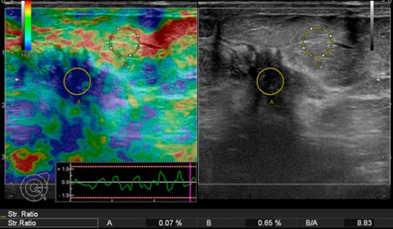
Background: Sonoelastography (SE) is introduced as a complementary technique for ultrasoungraohy (US) to evaluate breast lesions. This method is based on tissue strain in response to compression and decompression. The current study was designed to investigate the diagnostic performance of SE for differentiating between benign and malignant breast lesions

Methods: A total of 35 women with 45 breast lesions who were referred to a university affiliated hospital in Tehran were enrolled. All patients were visited and examined by a same radiologist. A five-point scale was applied for categorizing lesions in SE as malignant or benign. The results of US and SE were compared with histopathological results to calculate sensitivity and specificity of each mentioned techniques.

Results: Histopathological evaluations in 12 cases were in favor of malignancy, and the rest of cases were classified as benign. The sensitivity and specificity for US were 100% and 69.7%, respectively. On other hand, SE obtained a lower sensitivity (58.3%) and higher specificity (90.9%) in comparison with US.

Conclusions: simultaneous evaluation of suspicious breast lesions with both US and SE can have high sensitivity and specificity and prevent the unnecessary invasive interventions.

  • Article_DOI : doi.org/10.19187/abc.20141264-68
  • Author(s) : maryam rahmani
  • News Group : research,research article
  • News Code : 278486
مدیر سایت
Author:

مدیر سایت

Enter your desired term to search
Theme settings