Advanced Diagnostic & Interventional Radiology Research Center | 1H-MRSI and DWI for Non-invasive Grading of Brain Gliomas

Advanced Diagnostic & Interventional Radiology Research Center | 1H-MRSI and DWI for Non-invasive Grading of Brain Gliomas
| Jan 2 2026
logo

Advanced Diagnostic & Interventional Radiology Research Center

COVID-19 pandemic 

During the COVID-19 pandemic, the Radiology Research Center at Tehran University of Medical Sciences continued its research activities despite the challenges posed by the increased demand for CT scans of COVID-19 patients and the necessity of adhering to strict health protocols. This center played a crucial role in improving medical imaging techniques, optimizing diagnostic protocols, and advancing technologies related to CT scan image analysis.

Faculty members, researchers, and staff remained committed to ensuring the safety and well-being of healthcare professionals and patients while actively engaging in imaging data analysis, developing artificial intelligence algorithms for faster disease detection, publishing scientific articles, and presenting their findings at international conferences. These efforts aimed to enhance diagnostic accuracy, improve treatment processes, and alleviate pressure on healthcare systems.


Key achievements of the Radiology Research Center during the COVID-19 pandemic include:


✔️ Development and optimization of lung imaging protocols for faster and more accurate COVID-19 diagnosis
✔️ Implementation of artificial intelligence technologies for automated CT scan analysis and reduced diagnosis time
✔️ Publication of high-impact research articles on innovative imaging methods for COVID-19 patients
✔️ Participation in national and international projects focused on COVID-19 diagnosis and patient management

The center remains dedicated to advancing research in medical imaging and continues to contribute as a leading scientific institution in improving the quality of diagnostic and therapeutic services.


Some of the center's significant achievements during the pandemic include:


  • Release Date : Mar 17 2024 - 13:01
  • : 18
  • Study time : 1 minute(s)

Efficacy of 1H-MRSI and DWI for Non-invasive Grading of Brain Gliomas

1H-MRSI and DWI for Non-invasive Grading of Brain Gliomas {faces}

Background:

Distinguishing low-grade from high-grade gliomas can aid in optimal treatment planning and prognostication. Diffusion-weighted imaging (DWI) and magnetic resonance spectroscopy (MRS) have been applied in several studies for non-invasive glioma grading. However, these studies focused on limited aspects of these imaging techniques and used different study setups, resulting in occasionally inconsistent and incomparable conclusions in the literature

Objectives:

This study was designed to introduce the optimal imaging setup and the most reliable and applicable imaging parameters in glioma grading, using DWI and MRS.

Patients and Methods:

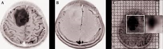
During this prospective study, using a 3T-MR scanner, 55 glioma patients underwent brain MRS with short, intermediate, and long echo times (TEs), as well as DWI using low, intermediate, and high b-values. Postoperatively, all of the specimens were graded pathologically using light microscopy.

Results:

We found that Max (Chol/Cr)/ Min (NAA/Cr) ((maximum choline to creatine ratio)/(minimum N-acetyl aspartate to creatine ratio)), followed by Max (Chol/ Cr), both in long-TE, were the most reliable metabolite ratios on MRS for accurate glioma grading. These had values for area under the curve (AUC) of 0.92 (P < 0.05) and 0.89 (P = 0.001), respectively, compared to conventional MR imaging (cMRI), which had an AUC of 0.83 (P < 0.05). DWI at maximal accuracy showed an AUC of 0.80 (P < 0.05).

Conclusion:

Max (Chol/Cr)/Min (NAA/Cr) in long-TE was the most reliable of all of the MRS parameters studied, while DWI showed no superiority over cMRI in glioma grading. No significant differences existed among the various b-values applied, or between the minimum and mean tumor apparent diffusion coefficient values used in DWI-based glioma grading

  • Article_DOI : doi.org/10.5812/iranjradiol.17817
  • Author(s) : mojtaba miri ,meysam mohseni
  • News Group : research,research article
  • News Code : 278422
مدیر سایت
Author:

مدیر سایت

Enter your desired term to search
Theme settings