Advanced Diagnostic & Interventional Radiology Research Center | Brain and Cervical MRI Abnormality Rates

Advanced Diagnostic & Interventional Radiology Research Center | Brain and Cervical MRI Abnormality Rates
| Aug 5 2026
logo

Advanced Diagnostic & Interventional Radiology Research Center

COVID-19 pandemic 

During the COVID-19 pandemic, the Radiology Research Center at Tehran University of Medical Sciences continued its research activities despite the challenges posed by the increased demand for CT scans of COVID-19 patients and the necessity of adhering to strict health protocols. This center played a crucial role in improving medical imaging techniques, optimizing diagnostic protocols, and advancing technologies related to CT scan image analysis.

Faculty members, researchers, and staff remained committed to ensuring the safety and well-being of healthcare professionals and patients while actively engaging in imaging data analysis, developing artificial intelligence algorithms for faster disease detection, publishing scientific articles, and presenting their findings at international conferences. These efforts aimed to enhance diagnostic accuracy, improve treatment processes, and alleviate pressure on healthcare systems.


Key achievements of the Radiology Research Center during the COVID-19 pandemic include:


✔️ Development and optimization of lung imaging protocols for faster and more accurate COVID-19 diagnosis
✔️ Implementation of artificial intelligence technologies for automated CT scan analysis and reduced diagnosis time
✔️ Publication of high-impact research articles on innovative imaging methods for COVID-19 patients
✔️ Participation in national and international projects focused on COVID-19 diagnosis and patient management

The center remains dedicated to advancing research in medical imaging and continues to contribute as a leading scientific institution in improving the quality of diagnostic and therapeutic services.


Some of the center's significant achievements during the pandemic include:


  • Release Date : Dec 20 2023 - 10:31
  • : 192
  • Study time : 1 minute(s)

Evaluation Of Brain And Cervical Mri Abnormality Rates In Patients With Systemic Lupus Erythematosus With Or Without Neurological Manifestations

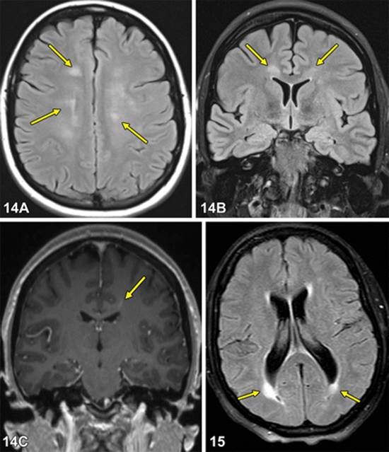
The aim of this study was to evaluate the prevalence and extent of brain and cervical cord MRI lesions of lupus patients.
Systemic Lupus Erythematosus With Or Without Neurological Manifestations {faces}

Background

Central nervous system (CNS) involvement has been observed in 14-80% of patients with systemic lupus erythematosus (SLE). Magnetic resonance imaging (MRI) is an appropriate method for evaluating CNS involvement in these patients. Clinical manifestations and MRI findings of CNS lupus should be differentiated from other mimicking diseases such as multiple sclerosis (MS).

Objectives

The aim of this study was to evaluate the prevalence and extent of brain and cervical cord MRI lesions of lupus patients. The relationship between neurological signs and symptoms and MRI findings were evaluated as well.

Patients and Methods

Fifty SLE patients who had been referred to the rheumatology clinic of our hospital within 2009 were included in a cross sectional study. All patients fulfilled the revised 1981 American College of Rheumatology (ACR) criteria for SLE. We evaluated the neurological signs and symptoms and brain and cervical MRI findings in these patients.

Results

Forty-one patients (82%) were female and nine (18%) were male. The mean age was 30.1 ± 9.3 years. Twenty eight (56%) patients had an abnormal brain MRI. No one showed any abnormality in the cervical MRI. The lesions in 20 patients were similar to demyelinative plaques. Seventeen patients with abnormal brain MRI were neurologically asymptomatic. There was only a significant relationship between neurological motor manifestations and brain MRI abnormal findings.

Conclusions

Unlike the brain, cervical MRI abnormality and especially asymptomatic cord involvement in MRI is quite rare in SLE patients. This finding may be helpful to differentiate SLE from other CNS disorders such as MS.

Keywords: Lupus Erythematosus Systemic, Brain, Magnetic Resonance Imaging, Neurologic Manifestations
  • Article_DOI : 10.5812/kmp.iranjradiol.17351065.3138
  • Author(s) : mohammad hossein harirchian,hazhir saberi
  • News Group : research,research article
  • News Code : 278571
مدیر سایت
Author:

مدیر سایت

Enter your desired term to search
Theme settings