Advanced Diagnostic & Interventional Radiology Research Center | Fetal intracranial hemorrhage and infarct

Advanced Diagnostic & Interventional Radiology Research Center | Fetal intracranial hemorrhage and infarct
| Aug 4 2026
logo

Advanced Diagnostic & Interventional Radiology Research Center

COVID-19 pandemic 

During the COVID-19 pandemic, the Radiology Research Center at Tehran University of Medical Sciences continued its research activities despite the challenges posed by the increased demand for CT scans of COVID-19 patients and the necessity of adhering to strict health protocols. This center played a crucial role in improving medical imaging techniques, optimizing diagnostic protocols, and advancing technologies related to CT scan image analysis.

Faculty members, researchers, and staff remained committed to ensuring the safety and well-being of healthcare professionals and patients while actively engaging in imaging data analysis, developing artificial intelligence algorithms for faster disease detection, publishing scientific articles, and presenting their findings at international conferences. These efforts aimed to enhance diagnostic accuracy, improve treatment processes, and alleviate pressure on healthcare systems.


Key achievements of the Radiology Research Center during the COVID-19 pandemic include:


✔️ Development and optimization of lung imaging protocols for faster and more accurate COVID-19 diagnosis
✔️ Implementation of artificial intelligence technologies for automated CT scan analysis and reduced diagnosis time
✔️ Publication of high-impact research articles on innovative imaging methods for COVID-19 patients
✔️ Participation in national and international projects focused on COVID-19 diagnosis and patient management

The center remains dedicated to advancing research in medical imaging and continues to contribute as a leading scientific institution in improving the quality of diagnostic and therapeutic services.


Some of the center's significant achievements during the pandemic include:


  • Release Date : Feb 22 2026 - 09:19
  • : 34
  • Study time : Less than one minute

Fetal intracranial hemorrhage and infarct: Main sonographic and MRI characteristics: A review article
Fetal intracranial hemorrhage and infarct {faces}

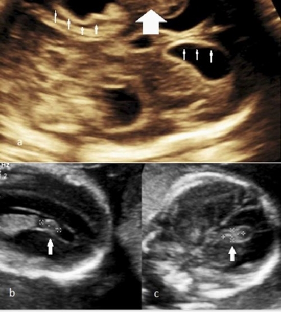
Early detection of fetal intracranial hemorrhage and infarct during pregnancy is crucial for preventing lethal and debilitating complications in neonatal life. Every radiologist must be aware of the imaging features of these conditions to refer patients to specialists. Sonographic and MRI features of fetal intracranial hemorrhage and infarct have been discussed in many previous articles. The aim of this article is to organize and categorize these findings into a practical guideline for improved application in diagnosing these diseases. The use of MRI sequences, such as DWI and multiplanar EPI should be developed for suspected prenatal infarct and intracranial hemorrhage and can serve as additional tools for early detection. In this review article, we first explain possible etiologic factors contributing to the development of fetal IVH and infarct. Then we discuss the different imaging features of these disorders on sonography and MRI separately, as well as their differential diagnosis. Finally, the mortality and morbidity associated with these two concerning fetal abnormalities will be addressed.

  • Article_DOI : 10.1016/j.eurox.2024.100351
  • Author(s) : behnaz moradi ,reihaneh mortazavi ardestani
  • News Group : research,research article
  • News Code : 316014
نفیسه سادات قوامی
Author:

نفیسه سادات قوامی

0 Comments for this article

comment

Post your comment:

متن درون تصویر را در جعبه متن زیر وارد نمائید *
Enter your desired term to search
Theme settings