Advanced Diagnostic & Interventional Radiology Research Center | Magnetic Resonance Imaging Features of Adenosis in the Breast

Advanced Diagnostic & Interventional Radiology Research Center | Magnetic Resonance Imaging Features of Adenosis in the Breast
| Jan 1 2026
logo

Advanced Diagnostic & Interventional Radiology Research Center

COVID-19 pandemic 

During the COVID-19 pandemic, the Radiology Research Center at Tehran University of Medical Sciences continued its research activities despite the challenges posed by the increased demand for CT scans of COVID-19 patients and the necessity of adhering to strict health protocols. This center played a crucial role in improving medical imaging techniques, optimizing diagnostic protocols, and advancing technologies related to CT scan image analysis.

Faculty members, researchers, and staff remained committed to ensuring the safety and well-being of healthcare professionals and patients while actively engaging in imaging data analysis, developing artificial intelligence algorithms for faster disease detection, publishing scientific articles, and presenting their findings at international conferences. These efforts aimed to enhance diagnostic accuracy, improve treatment processes, and alleviate pressure on healthcare systems.


Key achievements of the Radiology Research Center during the COVID-19 pandemic include:


✔️ Development and optimization of lung imaging protocols for faster and more accurate COVID-19 diagnosis
✔️ Implementation of artificial intelligence technologies for automated CT scan analysis and reduced diagnosis time
✔️ Publication of high-impact research articles on innovative imaging methods for COVID-19 patients
✔️ Participation in national and international projects focused on COVID-19 diagnosis and patient management

The center remains dedicated to advancing research in medical imaging and continues to contribute as a leading scientific institution in improving the quality of diagnostic and therapeutic services.


Some of the center's significant achievements during the pandemic include:


  • Release Date : Mar 3 2024 - 10:49
  • : 34
  • Study time : 1 minute(s)

Magnetic Resonance Imaging Features of Adenosis in the Breast

we aim to describe the features of breast adenosis lesions with suspicious or borderline findings on dynamic contrast-enhanced magnetic resonance imaging (DCE-MRI)

Magnetic Resonance Imaging Features of Adenosis in the Breast {faces}

Purpose

Adenosis lesions of the breast, including sclerosing adenosis and adenosis tumors, are a group of benign proliferative disorders that may mimic the features of malignancy on imaging. In this study, we aim to describe the features of breast adenosis lesions with suspicious or borderline findings on dynamic contrast-enhanced magnetic resonance imaging (DCE-MRI).

Methods

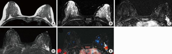
In our database, we identified 49 pathologically proven breast adenosis lesions for which the final assessment of the breast MRI report was classified as either category 4 (n=45) or category 5 (n=4), according to the Breast Imaging Reporting and Data System (BI-RADS) published by the American College of Radiology (ACR). The lesions had a final diagnosis of either pure adenosis (n=33, 67.3%) or mixed adenosis associated with other benign pathologies (n=16, 32.7%).

Results

Of the 49 adenosis lesions detected on DCE-MRI, 32 (65.3%) appeared as enhancing masses, 16 (32.7%) as nonmass enhancements, and one (2.1%) as a tiny enhancing focus. Analysis of the enhancing masses based on the ACR BI-RADS lexicon revealed that among the mass descriptors, the most common features were irregular shape in 12 (37.5%), noncircumscribed margin in 20 (62.5%), heterogeneous internal pattern in 16 (50.0%), rapid initial enhancement in 32 (100.0%), and wash-out delayed en-hancement pattern in 21 (65.6%). Of the 16 nonmass enhancing lesions, the most common descriptors included focal distribution in seven (43.8%), segmental distribution in six (37.5%), clumped internal pattern in nine (56.3%), rapid initial enhancement in 16 (100.0%), and wash-out delayed enhancement pattern in eight (50.0%).

Conclusion

Adenosis lesions of the breast may appear suspicious on breast MRI. Awareness of these suspi-cious-appearing features would be helpful in obviating unnecessary breast biopsies.

  • Article_DOI : 10.4048/jbc.2015.18.2.187
  • Author(s) : masoumeh gity ,elham taheri
  • News Group : research article,research
  • News Code : 278473
مدیر سایت
Author:

مدیر سایت

Enter your desired term to search
Theme settings