Advanced Diagnostic & Interventional Radiology Research Center | Massive hemothorax following CT-guided lung biopsy

Advanced Diagnostic & Interventional Radiology Research Center | Massive hemothorax following CT-guided lung biopsy
| Aug 3 2026
logo

Advanced Diagnostic & Interventional Radiology Research Center

COVID-19 pandemic 

During the COVID-19 pandemic, the Radiology Research Center at Tehran University of Medical Sciences continued its research activities despite the challenges posed by the increased demand for CT scans of COVID-19 patients and the necessity of adhering to strict health protocols. This center played a crucial role in improving medical imaging techniques, optimizing diagnostic protocols, and advancing technologies related to CT scan image analysis.

Faculty members, researchers, and staff remained committed to ensuring the safety and well-being of healthcare professionals and patients while actively engaging in imaging data analysis, developing artificial intelligence algorithms for faster disease detection, publishing scientific articles, and presenting their findings at international conferences. These efforts aimed to enhance diagnostic accuracy, improve treatment processes, and alleviate pressure on healthcare systems.


Key achievements of the Radiology Research Center during the COVID-19 pandemic include:


✔️ Development and optimization of lung imaging protocols for faster and more accurate COVID-19 diagnosis
✔️ Implementation of artificial intelligence technologies for automated CT scan analysis and reduced diagnosis time
✔️ Publication of high-impact research articles on innovative imaging methods for COVID-19 patients
✔️ Participation in national and international projects focused on COVID-19 diagnosis and patient management

The center remains dedicated to advancing research in medical imaging and continues to contribute as a leading scientific institution in improving the quality of diagnostic and therapeutic services.


Some of the center's significant achievements during the pandemic include:


  • Release Date : Jun 20 2026 - 09:50
  • : 13
  • Study time : Less than one minute

Massive hemothorax following CT-guided lung biopsy: A rare iatrogenic complication managed conservatively
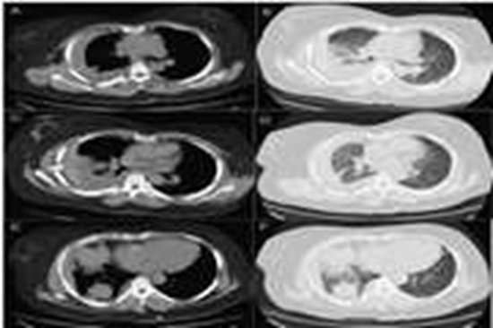
Massive hemothorax following CT-guided lung biopsy {faces}

CT-guided transthoracic lung biopsy is an essential diagnostic technique for evaluating pulmonary lesions. Although rare, major complications such as hemothorax can be life-threatening. We report the case of a 35-year-old woman who developed a rapidly enlarging right hemothorax several hours after CT-guided core needle biopsy of a pleura-abutting right lower lobe mass. There was no arterial extravasation, and chest tube drainage yielded approximately 1500 mL of dark, clotted blood, suggesting a venous or tumoral source. The patient remained hemodynamically stable and achieved complete recovery with conservative management. This case highlights that even massive post-biopsy hemothorax can be successfully treated non-operatively in stable patients by adhering to trauma-based management principles.

  • Article_DOI : 10.1016/j.tcr.2026.101303
  • Author(s) : bahman rasuli,ali forat yazdi
  • News Group : research,case report
  • News Code : 323921
نفیسه سادات قوامی
Author:

نفیسه سادات قوامی

0 Comments for this article

comment

Post your comment:

متن درون تصویر را در جعبه متن زیر وارد نمائید *
Enter your desired term to search
Theme settings