Advanced Diagnostic & Interventional Radiology Research Center | Distribution and Associated Anomalies Diagnosed by Prenatal Ultr

Advanced Diagnostic & Interventional Radiology Research Center | Distribution and Associated Anomalies Diagnosed by Prenatal Ultr
| Dec 13 2025
logo

Advanced Diagnostic & Interventional Radiology Research Center

COVID-19 pandemic 

During the COVID-19 pandemic, the Radiology Research Center at Tehran University of Medical Sciences continued its research activities despite the challenges posed by the increased demand for CT scans of COVID-19 patients and the necessity of adhering to strict health protocols. This center played a crucial role in improving medical imaging techniques, optimizing diagnostic protocols, and advancing technologies related to CT scan image analysis.

Faculty members, researchers, and staff remained committed to ensuring the safety and well-being of healthcare professionals and patients while actively engaging in imaging data analysis, developing artificial intelligence algorithms for faster disease detection, publishing scientific articles, and presenting their findings at international conferences. These efforts aimed to enhance diagnostic accuracy, improve treatment processes, and alleviate pressure on healthcare systems.


Key achievements of the Radiology Research Center during the COVID-19 pandemic include:


✔️ Development and optimization of lung imaging protocols for faster and more accurate COVID-19 diagnosis
✔️ Implementation of artificial intelligence technologies for automated CT scan analysis and reduced diagnosis time
✔️ Publication of high-impact research articles on innovative imaging methods for COVID-19 patients
✔️ Participation in national and international projects focused on COVID-19 diagnosis and patient management

The center remains dedicated to advancing research in medical imaging and continues to contribute as a leading scientific institution in improving the quality of diagnostic and therapeutic services.


Some of the center's significant achievements during the pandemic include:


  • Release Date : Apr 2 2024 - 13:31
  • : 28
  • Study time : 1 minute(s)

Neural Tube Defects: Distribution and Associated Anomalies Diagnosed by Prenatal Ultrasonography in Iranian Fetuses

Distribution by Prenatal Ultrasonography in Iranian Fetuses {faces}

Objectives: The current study aimed at providing detailed information about the distribution, associated anomalies, and syndromes in Iranian fetuses with neural tube defects (NTDs)
Methods: The current study was conducted in Yas Females’ referral and teaching hospital in 18 months from 2014 to 2016. All fetuses with a prenatally detected neural tube defect were included in the study. Neural tube defect characterization, gestational age, maternal reproductive factors, maternal risk factors, and associated anomalies were recorded.
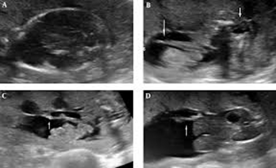
Results: Neural tube abnormalities were identified prenatally in 80 fetuses: 40 cases of ex/anencephaly, 22 cases of spina bifida, 13 cases of cephalocele and 5 cases of anencephaly /craniorachischisis. All the cases were detected before 21st week of gestation and 92.5% of the ex/anencephaly cases were diagnosed in the 1st trimester. Moreover, 40% of the open spina bifida cases in the current study were myelocele, while 75% of them referred only due to abnormal cranial findings. Incomplete consumption of folate was the most common associated risk factor (45%). Associated anomalies were recorded in 53 (66%) fetuses, with more prevalence in the fetuses with spina bifida (90%). Among the associated anomalies, central nervous system (CNS) anomalies were the most common type (26.26%). Chiari II was found in all the cases of open spina bifida and the ventriculomegaly rate was 30% in this group. Extremities anomalies and spine deformities were the 2nd and 3rd common associated findings, respectively. Limb-body-wall complex/amniotic band syndrome was the most common identified associated syndrome (6%).
Conclusions: Results of the current study confirmed the high prevalence of associated anomalies in neural tube defect cases and revealed the capability of detailed sonography to detect and define such abnormalities

  • Article_DOI : doi.org/10.5812/jogcr.64382
  • Author(s) : behnaz moradi ,fatemeh shakki katouli
  • News Group : research,research article
  • News Code : 278407
مدیر سایت
Author:

مدیر سایت

Enter your desired term to search
Theme settings