Advanced Diagnostic & Interventional Radiology Research Center | Pancreatic neuroendocrine tumors

Advanced Diagnostic & Interventional Radiology Research Center | Pancreatic neuroendocrine tumors
| Dec 12 2025
logo

Advanced Diagnostic & Interventional Radiology Research Center

COVID-19 pandemic 

During the COVID-19 pandemic, the Radiology Research Center at Tehran University of Medical Sciences continued its research activities despite the challenges posed by the increased demand for CT scans of COVID-19 patients and the necessity of adhering to strict health protocols. This center played a crucial role in improving medical imaging techniques, optimizing diagnostic protocols, and advancing technologies related to CT scan image analysis.

Faculty members, researchers, and staff remained committed to ensuring the safety and well-being of healthcare professionals and patients while actively engaging in imaging data analysis, developing artificial intelligence algorithms for faster disease detection, publishing scientific articles, and presenting their findings at international conferences. These efforts aimed to enhance diagnostic accuracy, improve treatment processes, and alleviate pressure on healthcare systems.


Key achievements of the Radiology Research Center during the COVID-19 pandemic include:


✔️ Development and optimization of lung imaging protocols for faster and more accurate COVID-19 diagnosis
✔️ Implementation of artificial intelligence technologies for automated CT scan analysis and reduced diagnosis time
✔️ Publication of high-impact research articles on innovative imaging methods for COVID-19 patients
✔️ Participation in national and international projects focused on COVID-19 diagnosis and patient management

The center remains dedicated to advancing research in medical imaging and continues to contribute as a leading scientific institution in improving the quality of diagnostic and therapeutic services.


Some of the center's significant achievements during the pandemic include:


  • Release Date : Jun 19 2024 - 06:45
  • : 35
  • Study time : 2 minute(s)

Pancreatic neuroendocrine tumors (pNETs): the predictive value of MDCT characteristics in the differentiation of histopathological grades

Pancreatic neuroendocrine tumors  {faces}

Purpose: To investigate the correlation between multiple detector computed tomography (MDCT) features of pancreatic neuroendocrine tumors (pNETs) and histopathologic grade and find valuable imaging criteria for grade prediction.

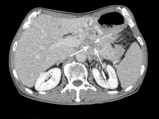
Material and methods: MDCT of 61 patients with 65 masses, which pNETs were approved histopathologically, underwent revision retrospectively. Each MDCT was evaluated for various radiologic characteristics. Absolute and relative (R: tumor/pancreas, D: tumor-pancreas) tumor enhancements were calculated in multiple post contrast phases.

Results: 61 patients [mean age = 50.70 ± 14.28 y/o and 30(49.2%) were male] were evaluated and classified into 2 groups histopathologically: G1: 32 (49.2%) and G2,3: 33 (50.8%). Significant relationships were observed between histopathologic tumor grade regarding age (p = 0.006), the longest tumor size (p = 0.006), presence of heterogeneity (p < 0.0001), hypodense foci in delayed phase (p = 0.004), lobulation (p = 0.002), vascular encasement (p < 0.0001), adjacent organ invasion (p = 0.01), presence (p < 0.0001) and number (0.02) of liver metastases, presence of lymphadenopathy with short axis of more than 10 mm (LAP) (p = 0.008), pathologic lymph node size (p = 0.004), relative (R and D) (p = 0.05 and 0.02, respectively), and percentage of arterial hyper-enhancing area (p = <0.0001). Tumor grades, however, had no significant relationship with gender, tumor location, tumor outline, calcification, cystic change, or pancreatic (PD) or biliary duct (BD) dilation (p = 0.21, 0.60, 0.05, 0.05 1, 0.10, and 0.51, respectively). Then, we suggested a novel imaging criteria consisting of six parameters (tumor size > 33 mm, relative (R) tumor enhancement in arterial phase ≤ 1.33, relative (D) tumor enhancement in arterial phase ≤ 16.5, percentage of arterial hyper-enhancing area ≤ 75%, vascular encasement, and lobulation), which specificity and accuracy of combination of all findings (6/6) for predicting G2,3 were 100% and 70.1%, respectively. The highest accuracy (84.21%) was seen in combinations of at least 4 of 6 findings, with 80.00% sensitivity, 87.5% specificity, 83.33% PPV, and 84.85% NPV.

  • Article_DOI : 10.1007/s00261-019-02372-x
  • Author(s) : niloofar ayoobi yazdi,faeze salahshour,mohammad-ali ataee
  • News Group : research,research article
  • News Code : 278291
مدیر سایت
Author:

مدیر سایت

Enter your desired term to search
Theme settings