Advanced Diagnostic & Interventional Radiology Research Center | Primary Hyperparathyroidism Misdiagnosed as Giant Cell Bone Tumo

Advanced Diagnostic & Interventional Radiology Research Center | Primary Hyperparathyroidism Misdiagnosed as Giant Cell Bone Tumo
| Jan 1 2026
logo

Advanced Diagnostic & Interventional Radiology Research Center

COVID-19 pandemic 

During the COVID-19 pandemic, the Radiology Research Center at Tehran University of Medical Sciences continued its research activities despite the challenges posed by the increased demand for CT scans of COVID-19 patients and the necessity of adhering to strict health protocols. This center played a crucial role in improving medical imaging techniques, optimizing diagnostic protocols, and advancing technologies related to CT scan image analysis.

Faculty members, researchers, and staff remained committed to ensuring the safety and well-being of healthcare professionals and patients while actively engaging in imaging data analysis, developing artificial intelligence algorithms for faster disease detection, publishing scientific articles, and presenting their findings at international conferences. These efforts aimed to enhance diagnostic accuracy, improve treatment processes, and alleviate pressure on healthcare systems.


Key achievements of the Radiology Research Center during the COVID-19 pandemic include:


✔️ Development and optimization of lung imaging protocols for faster and more accurate COVID-19 diagnosis
✔️ Implementation of artificial intelligence technologies for automated CT scan analysis and reduced diagnosis time
✔️ Publication of high-impact research articles on innovative imaging methods for COVID-19 patients
✔️ Participation in national and international projects focused on COVID-19 diagnosis and patient management

The center remains dedicated to advancing research in medical imaging and continues to contribute as a leading scientific institution in improving the quality of diagnostic and therapeutic services.


Some of the center's significant achievements during the pandemic include:


  • Release Date : Mar 16 2024 - 11:51
  • : 35
  • Study time : 1 minute(s)

Primary Hyperparathyroidism Misdiagnosed as Giant Cell Bone Tumor of Maxillary Sinus

Hyperparathyroidism Misdiagnosed as Giant Cell Bone Tumor of Maxillary Sinus {faces}

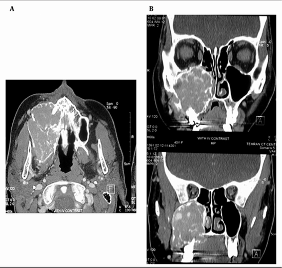
Primary hyperparathyroidism is an endocrine disorder recognized by hyperfunction of parathyroid gland, which can result in persistent bone absorption and brown tumor. Facial involvement of brown tumor is rare and usually involves the mandible. Giant cell tumor ( GCT) is an expansile osteolytic bone tumor which is very similar in clinical, radiological and histological features to brown tumor. Herein, we present a 35-year-old woman with an 11-month history of gradually swelling of the right maxilla and buccal spaces began during pregnancy two years ago. No other clinical or laboratory problems were detected. Postpartum CT scan demonstrated a lytic expansile multi-septated mass lesion containing enhancing areas, which initially described as GCT of the right maxillary sinus following surgery. Four months later, gradual progressive swelling of the bed of tumor was recurred and revised pathological slices were compatible with GCT. Regarding patient recent paresthesia, repeated laboratory tests were performed. Finally, according to laboratory results (elevation of serum calcium and parathyroid hormone), ultrasonographic findings and radioisotope scan (Sestamibi), probable parathyroid mass and brown tumor of maxilla was diagnosed. Pathology confirmed hyperplasia of right inferior parathyroid gland. Our case was thought-provoking due to its interesting clinical presentation and unusual presentation of brown tumor in parathyroid hyperplasia.

  • Article_DOI : 10.5812/iranjradiol.13260
  • Author(s) : leila aghaghazvini,hashem sharifian
  • News Group : research,research article
  • News Code : 278433
مدیر سایت
Author:

مدیر سایت

Enter your desired term to search
Theme settings