Advanced Diagnostic & Interventional Radiology Research Center | Segmentation of uterine using neighborhood information in uterin

Advanced Diagnostic & Interventional Radiology Research Center | Segmentation of uterine using neighborhood information in uterin
| Jan 2 2026
logo

Advanced Diagnostic & Interventional Radiology Research Center

COVID-19 pandemic 

During the COVID-19 pandemic, the Radiology Research Center at Tehran University of Medical Sciences continued its research activities despite the challenges posed by the increased demand for CT scans of COVID-19 patients and the necessity of adhering to strict health protocols. This center played a crucial role in improving medical imaging techniques, optimizing diagnostic protocols, and advancing technologies related to CT scan image analysis.

Faculty members, researchers, and staff remained committed to ensuring the safety and well-being of healthcare professionals and patients while actively engaging in imaging data analysis, developing artificial intelligence algorithms for faster disease detection, publishing scientific articles, and presenting their findings at international conferences. These efforts aimed to enhance diagnostic accuracy, improve treatment processes, and alleviate pressure on healthcare systems.


Key achievements of the Radiology Research Center during the COVID-19 pandemic include:


✔️ Development and optimization of lung imaging protocols for faster and more accurate COVID-19 diagnosis
✔️ Implementation of artificial intelligence technologies for automated CT scan analysis and reduced diagnosis time
✔️ Publication of high-impact research articles on innovative imaging methods for COVID-19 patients
✔️ Participation in national and international projects focused on COVID-19 diagnosis and patient management

The center remains dedicated to advancing research in medical imaging and continues to contribute as a leading scientific institution in improving the quality of diagnostic and therapeutic services.


Some of the center's significant achievements during the pandemic include:


  • Release Date : Jan 23 2024 - 14:08
  • : 38
  • Study time : 1 minute(s)

Segmentation of uterine using neighborhood information affected possibilistic fcm and gaussian mixture model in uterine fibroid patients MRI

In this paper, we proposed an automatic method for segmentation of uterus in MRIs

Segmentation of uterine {faces}

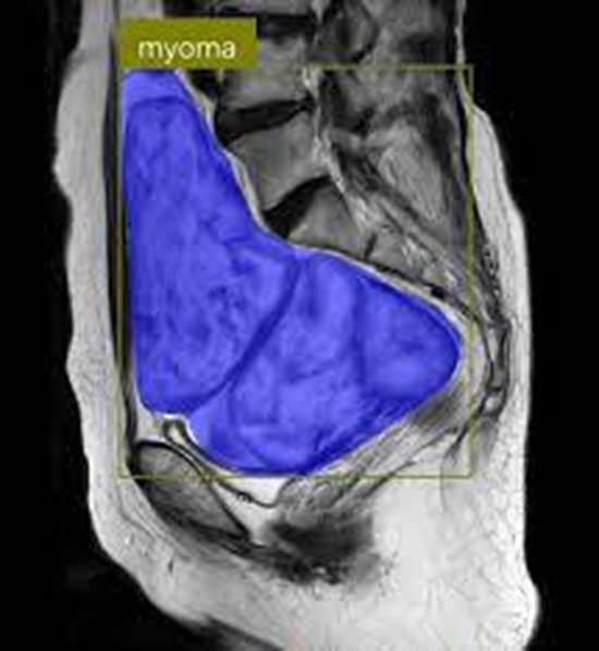
Uterine ¯broids are common tumors of female pelvis. Uterine volume measurement before and after surgery has an important role in predicting the outcome and later on in comparing with the result of the uterine ¯broid shrinkage surgery. Because of inhomogeneity and di®erent shapes and sizes of uterus and ¯broids, segmentation of uterus is a di±cult task. In this paper, using T1 and Enhanced-T1 MR images uterine is initially segmented using a new clustering algorithm named neighborhood information a®ected possibilistic fuzzy C-means (NIAPFCM). NIAPFCM uses membership, typicality and spatial neighborhood information to cluster each voxel. Finally, the redundant parts are removed by superimposing the segmented region of the T1-enhanced image over the registered T1 image. Gaussian mixture model (GMM) is applied to the extracted region histogram as a model for accurate tresholding. The results obtained using the proposed method are evaluated by comparing with manual segmentations using volume-based and distance-based metric methods. Also, the result of NIAPFCM is compared with fuzzy C-means (FCM) and possibilistic fuzzy C-means (PFCM) algorithms. We found this algorithm e±cient, which provides good and reliable results.


  • Article_DOI : 10.4015/S1016237214500100
  • Author(s) : hassan hashemi,alireza fallahi
  • News Group : research,research article,AI
  • News Code : 278497
مدیر سایت
Author:

مدیر سایت

Enter your desired term to search
Theme settings