Advanced Diagnostic & Interventional Radiology Research Center | Semiquantitative Dynamic Contrast-Enhanced MRI Accurate Classifi

Advanced Diagnostic & Interventional Radiology Research Center | Semiquantitative Dynamic Contrast-Enhanced MRI Accurate Classifi
| Jan 2 2026
logo

Advanced Diagnostic & Interventional Radiology Research Center

COVID-19 pandemic 

During the COVID-19 pandemic, the Radiology Research Center at Tehran University of Medical Sciences continued its research activities despite the challenges posed by the increased demand for CT scans of COVID-19 patients and the necessity of adhering to strict health protocols. This center played a crucial role in improving medical imaging techniques, optimizing diagnostic protocols, and advancing technologies related to CT scan image analysis.

Faculty members, researchers, and staff remained committed to ensuring the safety and well-being of healthcare professionals and patients while actively engaging in imaging data analysis, developing artificial intelligence algorithms for faster disease detection, publishing scientific articles, and presenting their findings at international conferences. These efforts aimed to enhance diagnostic accuracy, improve treatment processes, and alleviate pressure on healthcare systems.


Key achievements of the Radiology Research Center during the COVID-19 pandemic include:


✔️ Development and optimization of lung imaging protocols for faster and more accurate COVID-19 diagnosis
✔️ Implementation of artificial intelligence technologies for automated CT scan analysis and reduced diagnosis time
✔️ Publication of high-impact research articles on innovative imaging methods for COVID-19 patients
✔️ Participation in national and international projects focused on COVID-19 diagnosis and patient management

The center remains dedicated to advancing research in medical imaging and continues to contribute as a leading scientific institution in improving the quality of diagnostic and therapeutic services.


Some of the center's significant achievements during the pandemic include:


  • Release Date : Mar 17 2024 - 08:02
  • : 19
  • Study time : 1 minute(s)

Semiquantitative Dynamic Contrast-Enhanced MRI Accurate Classification of Complex Adnexal Masses

To identify the best dynamic contrast-enhanced (DCE) magnetic resonance imaging (MRI) descriptive parameters in predicting malignancy of complex ovarian masses, and develop an optimal decision tree for accurate classification of benign and malignant complex ovarian masses

 Dynamic Contrast-Enhanced MRI Accurate Classification ol Masses {faces}

Purpose: To identify the best dynamic contrast-enhanced (DCE) magnetic resonance imaging (MRI) descriptive parameters in predicting malignancy of complex ovarian masses, and develop an optimal decision tree for accurate classification of benign and malignant complex ovarian masses.

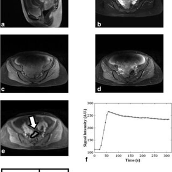
Materials and methods: Preoperative DCE-MR images of 55 sonographically indeterminate ovarian masses (27 benign and 28 malignant) were analyzed prospectively. Four descriptive parameters of the dynamic curve, namely, time-to-peak (TTP), wash-in-rate (WIR), relative signal intensity (SIrel ), and the initial area under the curve (IAUC60 ) were calculated on the normalized curves of specified regions-of-interest (ROIs). A two-tailed Student's t-test and two automated classifiers, linear discriminant analysis (LDA) and support vector machines (SVMs), were used to compare the performance of the mentioned parameters individually and in combination with each other.

Results: TTP (P = 6.15E-8) and WIR (P = 5.65E-5) parameters induced the highest sensitivity (89% for LDA, and 97% for SVM) and specificity (93% for LDA, and 100% for SVM), respectively. Regarding the high sensitivity of TTP and high specificity of WIR and through their combination, an accurate and simple decision-tree classifier was designed using the line equation obtained by LDA classification model. The proposed classifier achieved an accuracy of 89% and area under the ROC curve of 93%.

Conclusion: In this study an accurate decision-tree classifier based on a combination of TTP and WIR parameters was proposed, which provides a clinically flexible framework to aid radiologists/clinicians to reach a conclusive preoperative diagnosis and patient-specific therapy plan for distinguishing malignant from benign complex ovarian masses.

  • Article_DOI : 10.1002/jmri.25359
  • Author(s) : anahita fathi kazerooni, masoumeh gity
  • News Group : research article,research
  • News Code : 278430
مدیر سایت
Author:

مدیر سایت

Enter your desired term to search
Theme settings