Advanced Diagnostic & Interventional Radiology Research Center | Sonoelastography for skin evaluation in sclerodermic patients

Advanced Diagnostic & Interventional Radiology Research Center | Sonoelastography for skin evaluation in sclerodermic patients
| Dec 25 2025
logo

Advanced Diagnostic & Interventional Radiology Research Center

COVID-19 pandemic 

During the COVID-19 pandemic, the Radiology Research Center at Tehran University of Medical Sciences continued its research activities despite the challenges posed by the increased demand for CT scans of COVID-19 patients and the necessity of adhering to strict health protocols. This center played a crucial role in improving medical imaging techniques, optimizing diagnostic protocols, and advancing technologies related to CT scan image analysis.

Faculty members, researchers, and staff remained committed to ensuring the safety and well-being of healthcare professionals and patients while actively engaging in imaging data analysis, developing artificial intelligence algorithms for faster disease detection, publishing scientific articles, and presenting their findings at international conferences. These efforts aimed to enhance diagnostic accuracy, improve treatment processes, and alleviate pressure on healthcare systems.


Key achievements of the Radiology Research Center during the COVID-19 pandemic include:


✔️ Development and optimization of lung imaging protocols for faster and more accurate COVID-19 diagnosis
✔️ Implementation of artificial intelligence technologies for automated CT scan analysis and reduced diagnosis time
✔️ Publication of high-impact research articles on innovative imaging methods for COVID-19 patients
✔️ Participation in national and international projects focused on COVID-19 diagnosis and patient management

The center remains dedicated to advancing research in medical imaging and continues to contribute as a leading scientific institution in improving the quality of diagnostic and therapeutic services.


Some of the center's significant achievements during the pandemic include:


  • Release Date : Apr 20 2024 - 09:58
  • : 39
  • Study time : 1 minute(s)

Sonoelastography for skin evaluation in sclerodermic patients

Sonoelastography for skin evaluation in sclerodermic patients {faces}

Background: The objective of the study is to evaluate elastography ultrasound findings in patients with scleroderma (SS) and to clarify the effectiveness of elastosonography to differentiate scleroderma lesions from any skin lesion considering tissue elasticity.

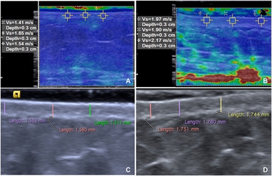
Methods: Thirty-six SS patients definite diagnosis of systemic sclerosis according to American College of Rheumatology criteria and 36 healthy subjects were enrolled. Volar aspect of the middle forearm and arm in addition to the dorsal aspect of the fingers were evaluated by sonoelastography. The RGB (red, green, blue) image is a three-dimensional matrix. A color image RGB is an M × N × 3 array of color pixels. The total pixels, total blue pixels, and blue index compared between SS cases and controls.

Results: Mean age of patients was 41.3 ± 10.3 years and mean age of controls was 39.8 ± 9.3 years. Mean-modified Rodnan skin score of the whole body was 11.9 and mean duration of disease was 6.2 years. Mean total blue pixels in the arm were significantly different between cases and controls. Mean total image pixels, total blue pixels, and blue index in the forearm were significantly different between cases and controls. Elastography findings in the finger were not significantly different between cases and controls.

Conclusions: Sonoelastography could be used for evaluating skin of forearm in sclerodermic cases which will be helpful for disease evaluation in clinical course

  • Article_DOI : 10.4103/ijpvm.IJPVM_43_18
  • Author(s) : arvin aryan,mahsa ghajarzadeh
  • News Group : research,research article
  • News Code : 278370
مدیر سایت
Author:

مدیر سایت

Enter your desired term to search
Theme settings