


The effect of superparamagnetic iron oxide nanoparticles surface engineering on relaxivity of magnetoliposome
The purpose of this work is evaluating the effect of ultra small superparamagnetic iron oxide nanoparticles (USPIONs) coatings on encapsulation efficiency in liposomes and cellular cytotoxicity assay
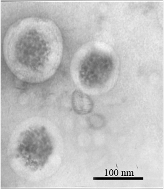
The purpose of this work is evaluating the effect of ultra small superparamagnetic iron oxide nanoparticles (USPIONs) coatings on encapsulation efficiency in liposomes and cellular cytotoxicity assay. Moreover, we assessed the effects of surface engineering on the relaxivity of magnetoliposome nanoparticles in order to create a targeted reagent for the intelligent diagnosis of cancers by MRI. For estimating the effect of nanoparticle coatings on encapsulation, several kinds of USPIONs coated by dextran, PEG5000 and citrate were used. All kinds of samples are monodispersed and below 100 ± 10 nm and the coatings of USPIONs have no significant effect on magnetoliposome diameter. The coating of USPIONs could have effect on percentage of encapsulation. The dextran coated USPIONs have more stability and quality accordingly the encapsulation increased up to 92%, then the magnetoliposome nano particles have been targeted by Herceptin and anti-HER2 VHH, separately. Over storage period of four weeks the resulting particles were stable and physico-chemical properties such as size and zetapotential did not show any significant changes. The relaxivity of contrast agents was measured using a 1.5 T MRI. The r2/r1 ratio was more than two for all samples which demonstrate the negative contrast enhancing of all SPION embedded specimens. The high ratio of r2/r1 as well as high r2 is the best combination of a negative contrast agent as it is obtained for pure magnetite. The value of r2/r1 for all other samples including Herceptin targeted magnetoliposome, anti-HER2 VHH targeted magnetoliposome and non-targeted magnetoliposome were between ~21 to ~28, which show the magnetite embedded samples have enough negative contrast to be detectable by MRI. Therefore the HER2 targeted magnetoliposomes are a good and stable candidate as contrast agents in clinical radiology and biomedical research with minimal cytotoxicity and biocompatibility effects. Copyright © 2016 John Wiley & Sons, Ltd.


Send to friends