Advanced Diagnostic & Interventional Radiology Research Center | Ultrasound Radio Frequency Time Series for Tissue Typing

Advanced Diagnostic & Interventional Radiology Research Center | Ultrasound Radio Frequency Time Series for Tissue Typing
| May 7 2026
logo

Advanced Diagnostic & Interventional Radiology Research Center

COVID-19 pandemic 

During the COVID-19 pandemic, the Radiology Research Center at Tehran University of Medical Sciences continued its research activities despite the challenges posed by the increased demand for CT scans of COVID-19 patients and the necessity of adhering to strict health protocols. This center played a crucial role in improving medical imaging techniques, optimizing diagnostic protocols, and advancing technologies related to CT scan image analysis.

Faculty members, researchers, and staff remained committed to ensuring the safety and well-being of healthcare professionals and patients while actively engaging in imaging data analysis, developing artificial intelligence algorithms for faster disease detection, publishing scientific articles, and presenting their findings at international conferences. These efforts aimed to enhance diagnostic accuracy, improve treatment processes, and alleviate pressure on healthcare systems.


Key achievements of the Radiology Research Center during the COVID-19 pandemic include:


✔️ Development and optimization of lung imaging protocols for faster and more accurate COVID-19 diagnosis
✔️ Implementation of artificial intelligence technologies for automated CT scan analysis and reduced diagnosis time
✔️ Publication of high-impact research articles on innovative imaging methods for COVID-19 patients
✔️ Participation in national and international projects focused on COVID-19 diagnosis and patient management

The center remains dedicated to advancing research in medical imaging and continues to contribute as a leading scientific institution in improving the quality of diagnostic and therapeutic services.


Some of the center's significant achievements during the pandemic include:


  • Release Date : Feb 22 2026 - 12:02
  • : 27
  • Study time : 1 minute(s)

Ultrasound Radio Frequency Time Series for Tissue Typing: Experiments on In-Vivo Breast Samples Using Texture-Optimized Features and Multi-Origin Method of Classification (MOMC)

Ultrasound Radio Frequency Time Series for Tissue Typing {faces}

Objectives: One of the most promising auxiliaries for screening breast cancer (BC) is ultrasound (US) radio-frequency (RF) time series. It has the superiority of not requiring any supplementary equipment over other methods. This article sought to propound a machine learning (ML) method for the automated categorization of breast lesions-categorized as benign, probably benign, suspicious, or malignant-using features extracted from the accumulated US RF time series.

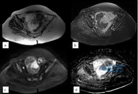
Methods: In this research, 220 data points of the categories as mentioned earlier, recorded from 118 patients, were analyzed. The RFTSBU dataset was registered by a SuperSonic Imagine Aixplorer® medical/research system fitted with a linear transducer. The expert radiologist manually selected regions of interest (ROIs) in B-mode images before extracting 283 features from each ROI in the ML approach, utilizing textural features such as Gabor filter (GF), gray-level co-occurrence matrix (GLCM), gray-level run-length matrix (GLRLM), gray-level size zone matrix (GLSZM), and gray-level dependence matrix (GLDM). Subsequently, the particle swarm optimization (PSO) narrowed the features to 131 highly effective ones. Ultimately, the features underwent classification using an innovative multi-origin method classification (MOMC), marking a significant leap in BC diagnosis.

Results: Employing 5-fold cross-validation, the study achieved notable accuracy rates of 98.57 ± 1.09%, 91.53 ± 0.89%, and 83.71 ± 1.30% for 2-, 3-, and 4-class classifications, respectively, using MOMC-SVM and MOMC-ensemble classifiers.

Conclusions: This research introduces an innovative ML-based approach to differentiate between diverse breast lesion types using in vivo US RF time series data. The findings underscore its efficacy in enhancing classification accuracy, promising significant strides in computer-aided diagnosis (CAD) for BC screening.

  • Article_DOI :
  • Author(s) :
  • News Group : research,research article
  • News Code : 316054
نفیسه سادات قوامی
Author:

نفیسه سادات قوامی

0 Comments for this article

comment

Post your comment:

متن درون تصویر را در جعبه متن زیر وارد نمائید *
Enter your desired term to search
Theme settings