


Evaluation of multimodal MR imaging for differentiating infiltrative versus reactive edema in brain gliomas
Objective: To determine the border of glial tumors by diffusion weighted imaging (DWI), apparent diffusion co-efficient (ADC), magnetic resonance spectroscopy (MRS) and perfusion brain MRI.
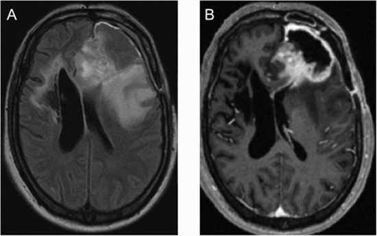
Patients and methods: Ten patients with brain gliomas were enrolled [mean age: 35.3 ± 13.2, range: 20-62]. Conventional MRI was performed for all patients. Besides, tumor mapping based on Choline (Cho)/Creatine (Cr) color map in MRS, perfusion and diffusion color maps, were gathered. Different tumoral and peritumoral regions [normal tissue, reactive edema, infiltrative edema, and tumor core] were defined. MRI criteria were evaluated in areas targeted for biopsy and histopathologic evaluation was determined.
Results: Tumor cell positive samples [one necrosis, 26 infiltrative and nine tumor cores] composed 36 (75%) of the 48 samples. Seven (19.4%) of the positive samples were interpreted as not tumor on MRI. Five were identified as reactive edema and two as normal tissue] [kappa: .67, p-value < .001]. Mean of ADC, median of N-acetylaspartate (NAA) and NAA/Cho were statistically different between positive and negative samples (p = .02 and p < .001, respectively). Mean ADC and median Cho/NAA were statistically different in missed tumor containing tissue presented as reactive edema compared to normal and correctly diagnosed reactive edema samples together (p-values < .05).
Conclusions: Multimodal MRI could define infiltrated borders of brain gliomas.


Send to friends