


Massive hemothorax following CT-guided lung biopsy: A rare iatrogenic complication managed conservatively
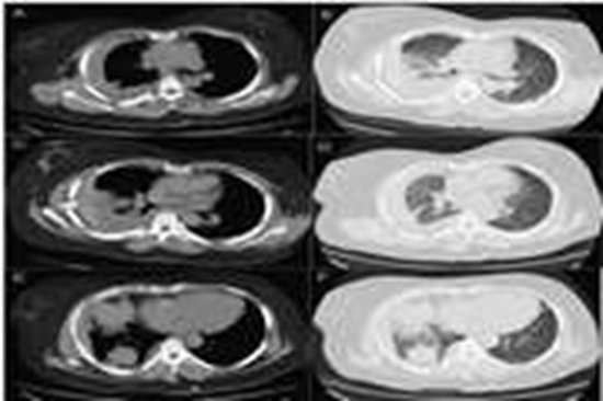
CT-guided transthoracic lung biopsy is an essential diagnostic technique for evaluating pulmonary lesions. Although rare, major complications such as hemothorax can be life-threatening. We report the case of a 35-year-old woman who developed a rapidly enlarging right hemothorax several hours after CT-guided core needle biopsy of a pleura-abutting right lower lobe mass. There was no arterial extravasation, and chest tube drainage yielded approximately 1500 mL of dark, clotted blood, suggesting a venous or tumoral source. The patient remained hemodynamically stable and achieved complete recovery with conservative management. This case highlights that even massive post-biopsy hemothorax can be successfully treated non-operatively in stable patients by adhering to trauma-based management principles.


comment