


Neuromuscular morphometric characteristics in low back pain with unilateral radiculopathy caused by disc herniation: An ultrasound imaging evaluation
Background: Little is known about the neuromuscular morphometric characteristics in patients with sciatica.
Objective: To evaluate the possible changes of nerve and muscle structures in patients with low back pain with unilateral radiculopathy due to lumbar disc herniation by ultrasound imaging.
Design: A case-control observational study.
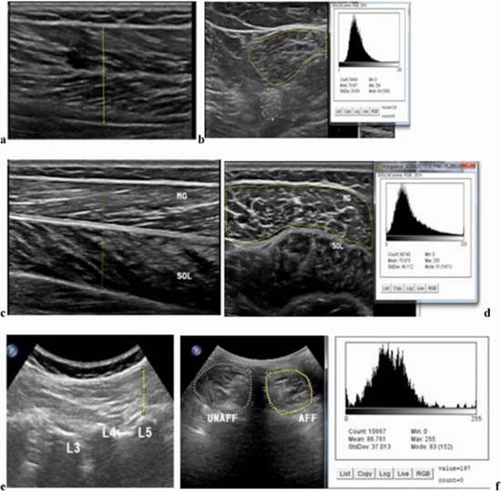
Methods: Forty individuals were divided into case (n = 20; low back pain with unilateral radiculopathy due to disc herniation), and healthy control groups (n = 20). The thickness of lumbar multifidus at L5 level, and of lower limb muscles (i.e., biceps femoris, medial gastrocnemius, and soleus) was measured during both rest and full contraction to calculate the rest/contraction ratio of these muscles. Additionally, the sciatic nerve cross-sectional area and the echogenicity of the nerve and muscles were measured based on ultrasound imaging. The association between severity of low back pain radiculopathy (i.e., pain and patients' perceived disability) and rest/contraction ratio was assessed.
Results: Patients with sciatica showed sciatic nerve enlargement, and different contraction ratios for multifidus (at L5)/ankle plantar flexors compared to the controls. The rest/contraction ratio for biceps femoris was similar between the two groups.
Conclusion: According to these findings, ultrasound imaging can be considered a useful tool to detect changes in the sciatic nerve and muscles due to disc herniation. Furthermore, regarding the observation of significant changes in muscle rest/contraction ratio in the multifidus and gastrosoleus, one might attribute these changes to the nerve root compression


Send to friends