


Perfusion weighted MRI patterns in neuropsychiatric systemic lupus erythematosus: a systematic review and meta-analysis
Background: Neuropsychiatric Systemic Lupus Erythematosus (NPSLE) is a complex manifestation of Systemic Lupus Erythematosus (SLE) characterized by a wide range of neurological and psychiatric symptoms. This study aims to elucidate the patterns of Perfusion-Weighted MRI (PWI) in NPSLE patients compared to SLE patients without neuropsychiatric manifestations (non-NPSLE) and healthy controls (HCs).
Material and methods: A systematic search was conducted in PubMed/Medline, Embase, Web of Science, and Scopus for studies utilizing PWI in NPSLE patients published through April 14, 2024. Cerebral blood flow (CBF) data from NPSLE, non-NPSLE patients, and HCs were extracted for meta-analysis, using standardized mean difference (SMD) as an estimate measure. For studies lacking sufficient data for inclusion, CBF, cerebral blood volume (CBV), and mean transit time (MTT) were reviewed qualitatively.
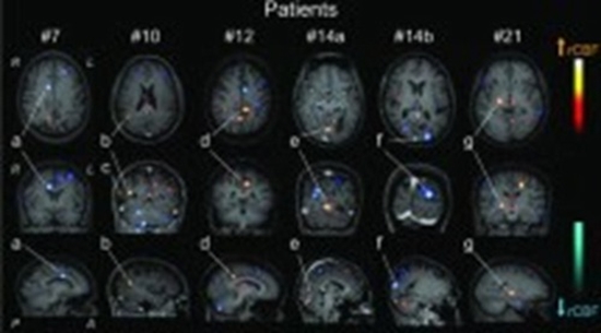
Results: Our review included eight observational studies employing PWI techniques, including dynamic susceptibility contrast (DSC) and arterial spin labeling (ASL). The meta-analysis of NPSLE compared to non-NPSLE incorporated four studies, encompassing 104 NPSLE patients and 90 non-NPSLE patients. The results revealed an SMD of -1.42 (95% CI: -2.85-0.00, I2: 94%) for CBF in NPSLE compared to non-NPSLE.
Conclusion: PWI reveals informative patterns of cerebral perfusion, showing a significant reduction in mean CBF in NPSLE patients compared to non-NPSLE patients. Our qualitative synthesis highlights these changes, particularly in the frontal and temporal lobes. However, the existing data exhibits considerable heterogeneity and limitations


comment