


Differentiating malignant from benign splenic lesions: a meta-analysis and pictorial review of imaging features
Objectives: Splenic lesions might exhibit overlapping imaging features, varying from benign entities like cysts and hemangiomas to malignancies such as lymphoma and angiosarcoma. This meta-analysis aims to delineate imaging characteristics that distinguish malignant from benign splenic lesions.
Methods: Adhering to PRISMA guidelines, we searched PubMed, Scopus, and Web of Science for studies on imaging features differentiating malignant from benign splenic lesions. We extracted data on splenic pathology and imaging characteristics and assessed the methodological quality via QUADAS-2. Odds ratio meta-analyses were performed using STATA (Version 17.0, Stata Corp, College Station, TX).
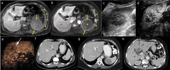
Results: Portal phase hypoenhancement, hypovascular enhancement pattern, diffusion restriction, and late phase hypoenhancement, with odds ratios above 10, highly indicate malignancy. Other features suggestive of malignancy include solid morphology, lymphadenopathy, presence of perisplenic fluid, arterial hypoenhancement, hypoechogenicity on ultrasound, splenomegaly, and presence of multiple lesions. In contrast, cystic morphology, hypervascular-washout and hypervascular-persistent pattern of enhancement, late phase hyperenhancement, anechogenicity on ultrasound, portal phase hyperenhancement, well-defined borders, and calcification are in favour of benign pathology.
Conclusion: The study underscores the critical role of contrast-enhanced and diffusion-weighted imaging in distinguishing malignant from benign splenic lesions, emphasizing the role of features like portal phase hypoenhancement and restricted diffusion in diagnosing malignancies. Additionally, the study emphasizes the value of contrast-enhanced ultrasound, which allows for the visualization of key contrast-enhancement patterns without the risk of ionizing radiation exposure.


ارسال نظر