


Quantitative Dynamic Contrast-Enhanced Magnetic Resonance Imaging and Positron Emission Tomography (PET) for Distinguishing Metastatic Lymph Nodes from Nonmetastatic Among Patients with Rectal Cancer: A Systematic Review and Meta-Analysis
Objective The objective of this research was to assess the proficiency of quantitative dynamic contrast-enhanced magnetic resonance imaging (QDCE-MRI) and positron emission tomography (PET) imaging in distinguishing between metastatic and nonmetastatic lymph nodes in cases of rectal carcinoma.
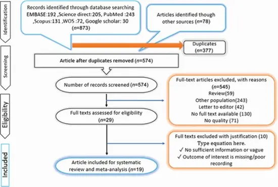
Method This meta-analysis was conducted following the Preferred Reporting Items for Systematic Reviews and Meta-Analyses standards. Two independent reviewers systematically searched databases including PubMed, Embase, Web of Science, and the Cochrane Library. The research took place in July 2022, with no restriction on the initial date of publication. For the analysis, we utilized Stata software (version 16.0), Review Manager (version 5.3), and the Open Meta-Analyst computational tool.
Results A total of 19 studies consisting of 1,451 patients were included in the current meta-analysis. The differences between metastatic and nonmetastatic lymph node parameters were significant by using short axis and Ktrans (6.9 ± 4 vs. 5.4 ± 0.5, 0.22 ± 0.1 vs. 0.14 ± 0.1, respectively). Contrast-enhanced MRI (CE-MRI) showed 73% sensitivity, 71% specificity, and 79% accuracy in detecting metastatic lymph nodes among rectal cancer patients based on six included studies ( n = 530). The overall sensitivity, specificity, and accuracy of QDCE-MRI using Ktrans was calculated to be 80, 79, and 80%, respectively. Furthermore, PET-computed tomography (CT) showed a sensitivity of 80%, specificity of 91%, and accuracy of 86% in distinguishing metastatic lymph nodes. Quality utility analysis showed that using CE-MRI, QDCE-MRI, and PET-CT would increase the posttest probability to 69, 73, and 85%, respectively.
Conclusion QDCE-MRI demonstrates a commendable sensitivity and specificity, but slightly overshadowed by the higher specificity of PET-CT at 91%, despite comparable sensitivities. However, the heterogeneity in PET-CT sensitivity across studies and its high specificity indicate variability that can influence clinical decision-making. Thus, combining these imaging techniques and perhaps newer methods like PET/MRI could enhance diagnostic accuracy, reduce variability, and improve patient management strategies in rectal cancer.


ارسال نظر