Management of intravenous contrast extravasations with ultrasonography: A case report
Extravasation of ionic and nonionic contrast materials is a well-recognized complication of contrast-enhanced imaging studies

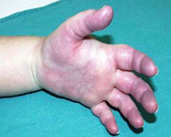
Extravasation of ionic and nonionic contrast materials is a well-recognized complication of contrast-enhanced imaging studies. Complications vary from minimal swelling to severe skin and subcutaneous ulceration, necrosis, and compartment syndrome. We report a case of Omnipaque (iohexol) extravasation in a 50-year-old man with erythema, blistering, and compartment syndrome who was treated medically but was not cured. Using gray scale ultrasonography, we determined the characteristics of the lesion, its distance from the skin, and its proximity to the vessels. We then determined the depth of the lesion, and then inserted the tip of the needle into the lesion. We also used ultrasonography in locations where extravasation was near an artery. After aspiration, the diameter of the lesion decreased significantly. The patient was cured by ultrasonography-guided aspiration from the extravasated site.


ارسال به دوستان