The ultrasonographic correlates of carpal tunnel syndrome in patients with normal electrodiagnostic tests
The aim of this study was to investigate the value of ultrasound imaging in diagnosing clinically suspicious patients with normal EDT findings

Purpose: The diagnosis of carpal tunnel syndrome (CTS) is established by electrodiagnostic testing (EDT). Nonetheless, in a portion of patients complaining of the typical signs and symptoms of CTS, the EDT is negative, and yet no paraclinical tool has been acknowledged for confirming the diagnosis. The aim of this study was to investigate the value of ultrasound imaging in diagnosing clinically suspicious patients with normal EDT findings.
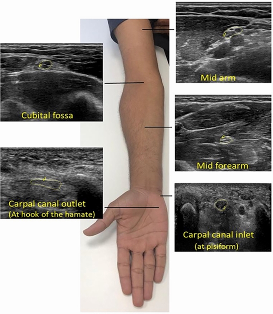
Materials and methods: Thirty-four patients, with clinical evidence of CTS but without abnormal findings on electromyography, and 41 healthy controls were enrolled. Ultrasonography was performed in all participants, and cross-sectional area (CSA), hypoechogenicity and hypervascularity of the median nerve were evaluated. Multivariate logistic regression analysis was used to formulate a prediction model for CTS.
Results: CSA of the median nerve in the wrist and wrist-to-forearm ratio were significantly higher in patients compared with controls. Patients had significantly higher hypoechogenicity [odds ratio (OR) 4.317; 95% confidence interval (CI) 1.23-15.11) and hypervascularity (OR 5.004,; 95% CI 1.02-21.15) in the median nerve. Clinical evidence of CTS was predicted using a model comprising three ultrasonographic determinant factors, including hypoechogenicity, hypervascularity and wrist CSA of the median nerve. The probability of clinical evidence of CTS in a person with one, two, or three ultrasonographic signs of CTS was estimated to be 35%, 70%, and 90%, respectively.
Conclusions: Ultrasound imaging is a useful technique in diagnosing CTS patients when EDT results are not confirmatory and the patient is suspected of having neuropathy.


ارسال به دوستان