


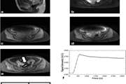
Magnetic Resonance Imaging Features of Adenosis in the Breast (vol 18, pg 187, 2015)
In this study, we aim to describe the features of breast adenosis lesions with suspicious or borderline findings on dynamic contrast-enhanced magnetic resonance imaging (DCE-MRI)
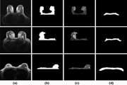
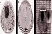
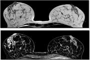
Localized-atlas-based segmentation of breast MRI in a decision-making framework
Breast-region segmentation is an important step for density estimation and Computer-Aided Diagnosis (CAD) systems in Magnetic Resonance Imaging (MRI)
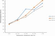
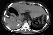
Semiquantitative Dynamic Contrast-Enhanced MRI Accurate Classification of Complex Adnexal Masses
To identify the best dynamic contrast-enhanced (DCE) magnetic resonance imaging (MRI) descriptive parameters in predicting malignancy of complex ovarian masses, and develop an optimal decision tree for accurate classification of benign and malignant complex ovarian masses
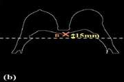
Diagnostic investigation of breast magnetic resonance imaging in malignant and benign mass lesions
Breast magnetic resonance imaging (BMRI) has been identified as a valuable modality in the diagnosis of breast cancer and monitoring the response to chemotherapy. The aim of this study was to evaluate the relative importance of different descriptors of breast masses in contrast-enhanced breast MRI
