Cortical Brain Mapping Using fmri
https://adir.tums.ac.ir//news/The-Human-Olfactory-System:-Cortical-Brain-Mapping-Using-fmri
The Human Olfactory System Background Functional magnetic resonance imaging (fMRI) is a non-invasive and convenient method of mapping brain activity associated with the human sensory systems. Among these systems there is a lack of data from olfactory studies, which could be attributed to technical difficulties in odor delivery during scanning. The current study took advantage of an olfactometer to evaluate brain activity during the odor-smelling process. Objectives This study aimed to investigat

Magnetic Resonance Imaging Features of Adenosis in the Breast
https://adir.tums.ac.ir//Research-Article/Magnetic-Resonance-Imaging-Features-of-Adenosis-in-the-Breast-(vol-18,-pg-187,-2015)
(vol 18, pg 187, 2015) In this study, we aim to describe the features of breast adenosis lesions with suspicious or borderline findings on dynamic contrast-enhanced magnetic resonance imaging (DCE-MRI) Purpose Adenosis lesions of the breast, including sclerosing adenosis and adenosis tumors, are a group of benign proliferative disorders that may mimic the features of malignancy on imaging. In this study, we aim to describe the features of breast adenosis lesions with suspicious or borderline fin

Localized-atlas-based segmentation of breast MRI in a decision-m
https://adir.tums.ac.ir//news/Localized-atlas-based-segmentation-of-breast-MRI-in-a-decision-making-framework
Localized atlas based segmentation of breast MRI in a decision making framework Breast-region segmentation is an important step for density estimation and Computer-Aided Diagnosis (CAD) systems in Magnetic Resonance Imaging (MRI) Breast-region segmentation is an important step for density estimation and Computer-Aided Diagnosis (CAD) systems in Magnetic Resonance Imaging (MRI). Detection of breast-chest wall boundary is often a difficult task due to similarity between gray-level values of fibrog

wrist Joint
https://adir.tums.ac.ir//news/Comparison-of-Quantitative-Assessment-of-BLADE-and-Isotropic-Three-Dimensional-Fast-Spin-Echo-Cube-(3D-T2-SPACE)-Sequences-with-Conventional-Protocols-of-wrist-Joint-at-3-Tesla-Magnetic-Resonance-Imaging
Comparison of Quantitative Assessment of BLADE and Isotropic Three Dimensional Fast Spin Echo Cube (3D T2 SPACE) Sequences with Conventional Protocols of at 3 Tesla Magnetic Resonance Imaging Background Magnetic resonance imaging (MRI) of the wrist joint is a useful method in the diagnosis of triangular fibrocartilage complex (TFCC), ligaments and tendons, peripheral nerves, cartilage and carpal tunnel syndrome. However, the evaluation of these small anatomical structures is a topic of investiga

Dynamic Contrast-Enhanced MRI Accurate Classification ol Masses
https://adir.tums.ac.ir//news/f-Complex-AdnexaSemiquantitative-Dynamic-Contrast-Enhanced-MRI-Accurate-Classification-ol-Masses
f Complex AdnexaSemiquantitative Dynamic Contrast Enhanced MRI Accurate Classification ol Masses To identify the best dynamic contrast-enhanced (DCE) magnetic resonance imaging (MRI) descriptive parameters in predicting malignancy of complex ovarian masses, and develop an optimal decision tree for accurate classification of benign and malignant complex ovarian masses Purpose To identify the best dynamic contrast-enhanced (DCE) magnetic resonance imaging (MRI) descriptive parameters in predicting

of breast magnetic resonance imaging in malignant and benign mas
https://adir.tums.ac.ir//article/Diagnostic-investigation-of-breast-magnetic-resonance-imaging-in-malignant-and-benign-mass-lesions
Diagnostic investigation s lesions Breast magnetic resonance imaging (BMRI) has been identified as a valuable modality in the diagnosis of breast cancer and monitoring the response to chemotherapy. The aim of this study was to evaluate the relative importance of different descriptors of breast masses in contrast-enhanced breast MRI Introduction Breast magnetic resonance imaging (BMRI) has been identified as a valuable modality in the diagnosis of breast cancer and monitoring the response to chem
Infratemporal fossa glial choristoma (Heterotopia)
https://adir.tums.ac.ir//article/Infratemporal-fossa-glial-choristoma-(Heterotopia):-A-rare-presentation--
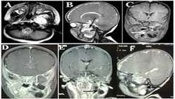
A rare presentation A 7-month-old girl was referred to the head and neck department with a history of gradual painless swelling over the left side of her face for 4 months. Physical examination revealed asymmetric appearance of the face, with a 120 × 70 mm non-tender, non-mobile, and non-pulsatile prominent soft tissue mass with a smooth contour in the left parotid and periauricular spaces. Neurological examination was unremarkable. On ultrasound examination, a solid cystic mass in the par

Hyperparathyroidism Misdiagnosed as Giant Cell Bone Tumor of Max
https://adir.tums.ac.ir//news/Primary-Hyperparathyroidism-Misdiagnosed-as-Giant-Cell-Bone-Tumor-of-Maxillary-Sinus
Primary illary Sinus Primary hyperparathyroidism is an endocrine disorder recognized by hyperfunction of parathyroid gland, which can result in persistent bone absorption and brown tumor. Facial involvement of brown tumor is rare and usually involves the mandible. Giant cell tumor ( GCT) is an expansile osteolytic bone tumor which is very similar in clinical, radiological and histological features to brown tumor. Herein, we present a 35-year-old woman with an 11-month history of gradually swelli

trunk paraspinal muscles before and after posterior lumbar surge
https://adir.tums.ac.ir//news/Cross-sectional-area-of-human-trunk-paraspinal-muscles-before-and-after-posterior-lumbar-surgery-using-magnetic-resonance-imaging
Cross sectional area of human ry using magnetic resonance imaging Iatrogenic injuries to paraspinal muscles during the posterior lumbar surgery (PLS) cause a reduction in their cross-sectional areas (CSAs) and contractile densities over time post-surgery. This study aims to quantify such alterations. Purpose Iatrogenic injuries to paraspinal muscles during the posterior lumbar surgery (PLS) cause a reduction in their cross-sectional areas (CSAs) and contractile densities over time post-surgery.
Whole-Body Bone Mineral Densitometry Measurements to Those From
https://adir.tums.ac.ir//news/Correlating-Whole–Body-Bone-Mineral-Densitometry-Measurements-to-Those-From-Local-Anatomical-Site/262283
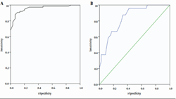
Correlating Whole Body Bone Mineral Densitometry Measurements to Those From Local Anatomical Site Background Using the same cutoff points for whole-body measurements as for site-specific measurements will result in underestimation of osteoporosis. Objectives We assessed the correlation between densitometry measurements for the whole body with those for the femur, lumbar spine, and forearm to evaluate the possibility of replacing site-specific values with whole-body measurements. Patients and Met