BI-RADS MRI descriptors distinguish non-mass lesions
https://adir.tums.ac.ir//article/Role-of-breast-MRI-BI-RADS-descriptors-in-discrimination-of-non-mass-enhancement-lesion:-A-systematic-review-&-meta-analysis
Role of breast MRI BI RADS descriptors in discrimination of non mass enhancement lesion A systematic review & meta analysis Objectives To evaluate the association of BI-RADS 5th edition distribution and type of enhancement descriptors with the malignancy of non-mass enhancement (NME) lesions. Methods Medline via PubMed, Scopus, Web of Science, ProQuest, and Embase databases were systematically searched from January 2013 to July 2022 for original studies, written in English, reporting the positiv
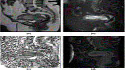
Advanced MRI model identifies uterine cancer site
https://adir.tums.ac.ir//news/Advanced-MRI-prediction-model-for-anatomical-site-identification-in-uterine-carcinoma:-enhancing-diagnostic-accuracy
Advanced MRI prediction model for anatomical site identification in uterine carcinoma enhancing diagnostic accuracy Aim The uterine carcinoma is the most commonly diagnosed malignancy in the female pelvis. Accurate identification of tumour origin is crucial for determining appropriate treatment approaches. This study aims to develop a prediction model using multiple MRI parameters to accurately diagnose uterine cancer with an indistinctive origin and those involving both the endometrium and cerv
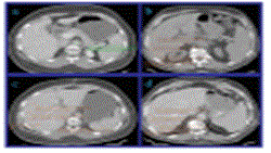
CT metrics reliability for liver fibrosis prediction
https://adir.tums.ac.ir//Research-Article/Reliability-assessment-of-CT-enhancement-rate-and-extracellular-volume-in-liver-fibrosis-prediction
Reliability assessment of CT enhancement rate and extracellular volume in liver fibrosis prediction Background Reliable, non-invasive evaluation of liver fibrosis is essential for early disease management. Computed tomography (CT)-based extracellular volume (ECV) fraction and portal venous phase enhancement rate (VP-ER) have shown potential in quantifying mild-to-moderate fibrosis. This study investigates the diagnostic performance of ECV and VP-ER in differentiating non-significant (F0-F1) from
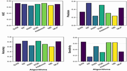
Machine learning accuracy for asthma diagnosis
https://adir.tums.ac.ir//news/Assessing-the-diagnostic-accuracy-of-machine-learning-algorithms-for-identification-of-asthma-in-United-States-adults-based-on-NHANES-dataset/293117
Assessing the diagnostic accuracy of machine learning algorithms for identification of asthma in United States adults based on NHANES dataset Asthma diagnosis poses challenges due to underreporting of symptoms, misdiagnoses, and limitations in existing diagnostic tests. Machine learning (ML) offers a promising avenue for addressing these challenges by leveraging demographic and clinical data. In this study, we aim to compare different ML diagnostic models and obtain the most valuable features fo

Pheochromocytoma in a patient with heterotaxy syndrome
https://adir.tums.ac.ir//news/Pheochromocytoma-in-a-patient-with-heterotaxy-syndrome:-a-case-report
a case report Background Heterotaxy syndrome is a rare congenital condition characterized by abnormal arrangement of thoracoabdominal organs, often associated with complex cardiac and splenic anomalies. Pheochromocytoma is a rare neuroendocrine tumor that overproduces catecholamines, leading to various complications. The co-occurrence of heterotaxy syndrome and pheochromocytoma has not been previously reported. This case report presents a unique finding of pheochromocytoma in a patient with hete

Hypofractionated vs. standard therapy for cervical cancer
https://adir.tums.ac.ir//article/Hypofractionated-versus-standard-chemoradiotherapy-in-the-definitive-treatment-of-uterine-cervix-cancer:-interim-results-of-a-randomized-controlled-clinical-trial
Hypofractionated versus standard chemoradiotherapy in the definitive treatment of uterine cervix cancer interim results of a randomized controlled clinical trial Purpose Concurrent chemoradiation has been the mainstay of treatment for cervix cancer. We aimed to evaluate the non-inferiority of hypofractionated chemoradiation. Methods This study was designed as a phase 2, 11 randomized, investigator-blinded, controlled, non-inferiority trial and we report the interim results after 50% accrual. Cer
.png)
Ultrasonographic Assessment of Intima-Media Thickness
https://adir.tums.ac.ir//Research-Article/Ultrasonographic-Assessment-of-Intima-Media-Thickness-(IMT),-as-a-Surrogate-of-Future-Atherosclerosis-and-Cardiac-Diseases-in-Patients-with-Hashimoto’s-Thyroiditis-and-Concomitant-Celiac-Disease:-A-Case-Control-Study
Ultrasonographic Assessment of Intima Media Thickness (IMT), as a Surrogate of Future Atherosclerosis and Cardiac Diseases in Patients with Hashimoto’s Thyroiditis and Concomitant Celiac Disease A Case Control Study Purpose Intima-Media Thickness (IMT), which is frequently evaluated by B-mode ultrasound study, has been proved to be a reliable surrogate marker for atherosclerosis progression and a predictor of upcoming cardiovascular risks. Sonographic changes of intima-media thickness of carotid
.png)
CADe and segmentation for breast cancer MRI
https://adir.tums.ac.ir//article/Computer–Aided-Detection-(CADe)-and-Segmentation-Methods-for-Breast-Cancer-Using-Magnetic-Resonance-Imaging-(MRI)
Computer-Aided Detection (CADe) and Segmentation Methods for Breast Cancer Using Magnetic Resonance Imaging (MRI) Breast cancer continues to be a major health concern, and early detection is vital for enhancing survival rates. Magnetic resonance imaging (MRI) is a key tool due to its substantial sensitivity for invasive breast cancers. Computer-aided detection (CADe) systems enhance the effectiveness of MRI by identifying potential lesions, aiding radiologists in focusing on areas of interest, e

Sonographic skin features and shear wave elastography
https://adir.tums.ac.ir//news/Sonographic-skin-features-and-shear-wave-elastography-in-distinguishing-active-from-inactive-morphea-lesions:-A-case–control-study/292774
in distinguishing active from inactive morphea lesions A case control study Morphea (localized scleroderma) is a connective tissue disorder characterized by dermal stiffness, thickening, erythema, and pigmentation abnormalities, often varying with the degree of inflammation and fibrosis.1 Precise lesion activity is crucial for tailoring optimal treatment plans. Traditional diagnostic methods like skin biopsy are invasive and may not effectively capture the dynamic progression of the disease.2 No

Expression of cytokeratin 19 in prostatic adenocarcinoma
https://adir.tums.ac.ir//article/Expression-of-cytokeratin-19-in-prostatic-adenocarcinoma:-a-systematic-review
a systematic review Background Cytokeratins are intracellular proteins known as diagnostic biomarkers or prognostic factors for certain cancers. Cytokeratin 19 (CK-19) expression has been proven to have prognostic value for some cancers, but its relationship with others, such as prostate cancer (PCa), remains unclear. This systematic review article aimed to examine the relationship between CK-19 expression and prostate adenocarcinoma (PAC). Methods To include the eligible studies that detected C