chest CT features as a predictive tool for COVID-19
https://adir.tums.ac.ir//news/Clinical-and-chest-CT-features-as-a-predictive-tool-for-COVID–19-clinical-progress:-introducing-a-novel-semi–quantitative-scoring-system/270751
Clinical and chest CT features as a predictive tool for COVID 19 clinical progress introducing a novel semi quantitative scoring system Objective Proposing a scoring tool to predict COVID-19 patients' outcomes based on initially assessed clinical and CT features. Methods All patients, who were referred to a tertiary-university hospital respiratory triage (March 27-April 26, 2020), were highly clinically suggestive for COVID-19 and had undergone a chest CT scan were included. Those with positive
.png)
dentate nucleus in multiple sclerosis patients
https://adir.tums.ac.ir//news/Evaluation-of-the-effect-of-multiple-linear-gadolinium–based-contrast-agent-exposures-on-the-signal-intensity-of-the-dentate-nucleus-in-multiple-sclerosis-patients/270749
Evaluation of the effect of multiple linear gadolinium based contrast agent exposures on the signal intensity of the Background Magnetic resonance imaging (MRI) with gadolinium (GAD)-based contrast agents has been the imaging modality of choice for early detection and monitoring of multiple sclerosis (MS) patients. Objectives This study aimed to assess the effect of multiple injections of linear GAD-based contrast agents on the signal intensity of the dentate nucleus (DN) in MS patients. Patie

Pancreatic neuroendocrine tumor presenting
https://adir.tums.ac.ir//news/Pancreatic-neuroendocrine-tumor-presenting-as-a-diffuse-pancreatic-enlargement,-case-report-and-review-of-literature
as a diffuse pancreatic enlargement, case report and review of literature Pancreatic neuroendocrine tumors are rare neoplasms that comprise 1–2% of all pancreatic tumors. However, they are the second most common solid pancreatic neoplasms. They have a wide range of imaging appearances and they can show common to very rare imaging presentations. Most of the time they are solitary well-marginated enhancing solid mass arising in a certain aspect of the pancreas. We present a case report of a

Outcome in Intensive Care Unit-Admitted COVID-19 Patients
https://adir.tums.ac.ir//Research-Article/Factors-Predicting-Outcome-in-Intensive-Care-Unit-Admitted-COVID-19-Patients:-Using-Clinical,-Laboratory,-and-Radiologic-Characteristics
Factors Predicting Outcome in Intensive Care Unit Admitted COVID 19 Patients Using Clinical, Laboratory, and Radiologic Characteristics Purpose. To investigate the factors contributing to mortality in coronavirus disease 2019 (COVID-19) patients admitted in the intensive care unit (ICU) and design a model to predict the mortality rate. Method. We retrospectively evaluated the medical records and CT images of the ICU-admitted COVID-19 patients who had an on-admission chest CT scan. We analyzed th
.png)
Isolated duodenal ischemia of unknown etiology
https://adir.tums.ac.ir//article/Isolated-duodenal-ischemia-of-unknown-etiology:-a-case-report
a case report Background Acute abdomen is among the most common presentations observed in clinical practice. The present study describes a patient with isolated duodenal ischemia as an extremely rare etiology of acute abdomen. Case presentation A 79-year-old male with acute abdominal pain, nausea, and vomiting presented to the emergency department of our hospital. He was diagnosed with myelodysplastic syndrome 7 years ago, for which he took thalidomide and erythropoietin as the main medications.

cerebral oxygen extraction fraction in MRI-negative refractory
https://adir.tums.ac.ir//news/An-interictal-measurement-of-cerebral-oxygen-extraction-fraction-in-MRI-negative-refractory-epilepsy-using-quantitative-susceptibility-mapping
An interictal measurement of cerebral oxygen extraction fraction in MRI negative refractory epilepsy using quantitative susceptibility mapping Purpose Oxygen extraction fraction (OEF) can be a factor to identify brain tissue's disability in epileptic patients. This study aimed to assess the OEF's level measurement in refractory epileptic patients (REPs) using a quantitative susceptibility mapping (QSM) method and to determine whether the OEF parameters change Methods QSM-OEF maps of 26 REPs and

Sparing Contralateral Fibrotic Lung
https://adir.tums.ac.ir//article/COVID-19:-Unilateral-Involvement-of-Transplanted-Lung,-Sparing-Contralateral-Fibrotic-Lung
COVID 19 Unilateral Involvement of Transplanted Lung, Introduction Organ transplant recipients might be more likely to develop COVID-19, as they receive long-term immunosuppressives and have comorbidities. Case Presentation Herein, we reported the case of a 32-year-old man with unilateral lung transplantation due to unclassifiable lung fibrosis on pathologic evaluation who presented with cough, fever, and headache. After evaluation with RT-PCR test and chest CT scan, COVID-19 in the previously t
.jpg)
Anthracosis, a Distinct Cause of Vocal Fold Paralysis
https://adir.tums.ac.ir//case-report/Anthracosis,-a-Distinct-Cause-of-Vocal-Fold-Paralysis:-Case-Series
Case Series Vocal fold paralysis (VFP) can happen in various conditions due to mediastinal LADs, however no study has proposed anthracosis as an etiology. Here we discussed the chest CT features of anthracosis related LADs causing VFP. Among 41 cases of pulmonary anthracosis, 10 had VFP that all were presented with hoarseness. The paralysis was unilateral (left side) in all cases. Extra-nodal infiltration and conglomeration of lymph nodes were significantly higher in patients with paralysis. Lef

Automated detection of pneumonia cases using deep transfer
https://adir.tums.ac.ir//news/Automated-detection-of-pneumonia-cases-using-deep-transfer-learning-with-paediatric-chest-X-ray-images
learning with paediatric chest X ray images Objective Pneumonia is a lung infection and causes the inflammation of the small air sacs (Alveoli) in one or both lungs. Proper and faster diagnosis of pneumonia at an early stage is imperative for optimal patient care. Currently, chest X-ray is considered as the best imaging modality for diagnosing pneumonia. However, the interpretation of chest X-ray images is challenging. To this end, we aimed to use an automated convolutional neural network-based
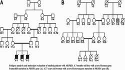
Identification of Two Novel Mutations in PKHD1 Gene
https://adir.tums.ac.ir//Research-Article/Identification-of-Two-Novel-Mutations-in-PKHD1-Gene-from-Two-Families-with-Polycystic-Kidney-Disease-by-Whole-Exome-Sequencing
from Two Families with Polycystic Kidney Disease by Whole Exome Sequencing Background Polycystic kidney disease (PKD) is an autosomal recessive disorder resulting from mutations in the PKHD1 gene on chromosome 6 (6p12), a large gene spanning 470 kb of genomic DNA. Objective The aim of the present study was to report newly identified mutations in the PKHD1 gene in two Iranian families with PKD. Materials and Methods Genetic alterations of a 3-month-old boy and a 27-year-old girl with PKD were eva