لیست اخبار صفحه :50 Proven Aspergillus flavus pulmonary aspergillosis in a COVID 19 patient: A case report and review of the literature 1403/04/18 - 11:26 Factors Predicting Outcome in Intensive Care Unit Admitted COVID 19 Patients: Using Clinical, Laboratory, and Radiologic Characteristics 1403/04/18 - 11:22 Isolated duodenal ischemia of unknown etiology: a case report 1403/04/18 - 11:18 An interictal measurement of cerebral oxygen extraction fraction in MRI negative refractory epilepsy using quantitative susceptibility mapping 1403/04/18 - 11:11 COVID 19: Unilateral Involvement of Transplanted Lung, Sparing Contralateral Fibrotic Lung 1403/04/18 - 11:01 Anthracosis, a Distinct Cause of Vocal Fold Paralysis: Case Series 1403/04/18 - 10:55 Automated detection of pneumonia cases using deep transfer learning with paediatric chest X ray images 1403/04/18 - 08:28 Identification of Two Novel Mutations in PKHD1 Gene from Two Families with Polycystic Kidney Disease by Whole Exome Sequencing 1403/04/18 - 08:23 Correction to: Predictors of the chest CT score in COVID 19 patients: a cross sectional study (Virology Journal, (2021), 18, 1, (225), 10.1186/s12985/021/01699/6) 1403/04/18 - 08:03 Daytime Hypersomnolence in COVID 19: A Case Report and Literature Review 1403/04/18 - 07:48 45 46 47 48 49 50 51 52 53 54 55
Proven Aspergillus flavus pulmonary aspergillosis in a COVID 19 patient: A case report and review of the literature 1403/04/18 - 11:26 Factors Predicting Outcome in Intensive Care Unit Admitted COVID 19 Patients: Using Clinical, Laboratory, and Radiologic Characteristics 1403/04/18 - 11:22 Isolated duodenal ischemia of unknown etiology: a case report 1403/04/18 - 11:18 An interictal measurement of cerebral oxygen extraction fraction in MRI negative refractory epilepsy using quantitative susceptibility mapping 1403/04/18 - 11:11 COVID 19: Unilateral Involvement of Transplanted Lung, Sparing Contralateral Fibrotic Lung 1403/04/18 - 11:01 Anthracosis, a Distinct Cause of Vocal Fold Paralysis: Case Series 1403/04/18 - 10:55 Automated detection of pneumonia cases using deep transfer learning with paediatric chest X ray images 1403/04/18 - 08:28 Identification of Two Novel Mutations in PKHD1 Gene from Two Families with Polycystic Kidney Disease by Whole Exome Sequencing 1403/04/18 - 08:23 Correction to: Predictors of the chest CT score in COVID 19 patients: a cross sectional study (Virology Journal, (2021), 18, 1, (225), 10.1186/s12985/021/01699/6) 1403/04/18 - 08:03 Daytime Hypersomnolence in COVID 19: A Case Report and Literature Review 1403/04/18 - 07:48
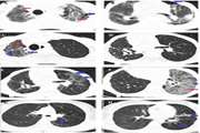
Proven Aspergillus flavus pulmonary aspergillosis in a COVID 19 patient: A case report and review of the literature 1403/04/18 - 11:26
Factors Predicting Outcome in Intensive Care Unit Admitted COVID 19 Patients: Using Clinical, Laboratory, and Radiologic Characteristics 1403/04/18 - 11:22
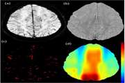
An interictal measurement of cerebral oxygen extraction fraction in MRI negative refractory epilepsy using quantitative susceptibility mapping 1403/04/18 - 11:11
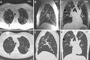
COVID 19: Unilateral Involvement of Transplanted Lung, Sparing Contralateral Fibrotic Lung 1403/04/18 - 11:01
Automated detection of pneumonia cases using deep transfer learning with paediatric chest X ray images 1403/04/18 - 08:28
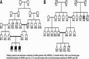
Identification of Two Novel Mutations in PKHD1 Gene from Two Families with Polycystic Kidney Disease by Whole Exome Sequencing 1403/04/18 - 08:23
Correction to: Predictors of the chest CT score in COVID 19 patients: a cross sectional study (Virology Journal, (2021), 18, 1, (225), 10.1186/s12985/021/01699/6) 1403/04/18 - 08:03


.png)
.png)
.jpg)
.png)